Gram Staining - Introduction, Purpose, Reagent, Principle, Procedure, Result
Introduction, Purpose of Gram Staining
Gram staining is a differential staining technique widely used to separate bacteria into two major groups- gram-positive bacteria and gram-negative bacteria. It is an essential method used for the classification and differentiation of bacteria.
Reagents for Gram Staining
The reagents used for gram staining include:
Primary stain: Crystal violet
Mordant: Gram's Iodine
Decolorizing agent: Ethyl alcohol (95%)
Counterstain: Safranin
Principle of Gram Staining
Bacterial cells contain pores through which stains can enter and give a prominent color, making their morphology and arrangement visible.
Primary stain
Crystal violet is used as a primary stain for gram staining. This stain colors all cells purple.
Mordant
Gram's Iodine serves as a mordant for gram staining that binds firmly with the primary stain i.e. crystal violet present resulting in the formation of crystal violet - Iodine (CV-I) complex. This complex intensifies the color of the stain and cells appear purple-black.
In cases of gram-positive organisms, the CV-I complex binds with the magnesium-ribonucleic acid of the cell wall forming magnesium-ribonucleic acid-crystal violet-Iodine complex (Mg-RNA-CV-I). Since Mg-RNA-CV-I is larger than just CV-I, it is more difficult to remove from bacterial cells.
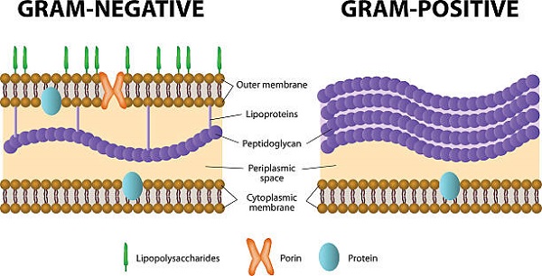
Fig: Bacterial cell wall composition (Source: iStock)
Decolorizing agent
The decolorizing agent ethyl alcohol works both as a protein-dehydrating agent and a lipid solvent in gram staining. In gram-positive microorganisms, the cell walls have low lipid concentration while in gram-negative microorganisms, the cell walls have high lipid concentration.
When ethyl alcohol is used, it dissolves the lipid present in the bacterial cell walls resulting in the formation of new cell wall pores. Such pores are larger in gram-negative bacteria than in Gram-positive bacteria, which suffer minute pores, due to high cell wall lipid concentration in Gram-negative microorganisms.
Moreover, small pores of gram-positive bacteria are closed off due to dehydrating effect of alcohol. But in the case of gram-negative bacteria, pores are not able to be closed off by alcohol's dehydrating effect. As a result, the CV-I complex escapes from gram-negative bacteria while the Mg-RNA-CV-I complex remains in gram-positive microorganisms.
Counterstain
Safranin is the dye of choice in the counterstaining procedure of gram staining. The decolorized cells, that have lost the CV-I complex absorb the counterstain changing its color to red.
Gram-positive bacteria still hold Mg-RNA-CV-I complex from the primary stain during the decolorizing procedure and are unable to take in the safranin that has been used as a counterstain. Thus, they appear purple in color.
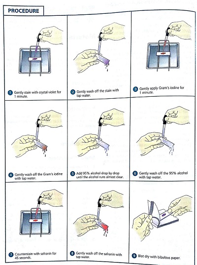
Fig: Gram staining procedure (Source: Otosection)
Procedure for Gram Staining
The procedure followed for gram staining is as follows:
Take a clean, grease-free slide
Prepare a bacterial smear of the test organism
Allow the smear to air dry
Heat fix the dried smear by quickly passing the smear through the bunsen burner 2-3 times
Flood the smear with primary stain (crystal violet) for 30 seconds to 1 minute
Wash the smear with slow-running water
Flood the smear with gram's iodine (mordant) for 1 minute
Wash the smear with slow-running water
Pour decolorizing agent (95% ethyl alcohol) on the smear drop by drop for 10-20 seconds
* the purple color of crystal violet is removed during this procedure
Wash the smear with slow-running water
Flood with counterstain (safranin) for 45 seconds
Gently tap the slide with blotting paper to remove the remaining water/dye
Examine under a microscope (oil immersion at 100X)
Heat fixation
Heat fixation is done in most staining procedures (except capsule staining, and negative staining) because, during the washing steps, the microbial smear could wash away. During heat fixation, the bacterial proteins are coagulated and fixed to the glass slide surface.
It is done by rapidly passing the air-dried smear 2-3 times over a bunsen burner's flame.
* Instead of heat fixation, several methods of heat fixation have been recognized. One such commonly used procedure is the alcohol-based fixative. Ethanol or methanol can also be used as they also function by precipitating proteins.
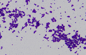
Fig: Gram-positive Staphylococcus (Source: ResearchGate)
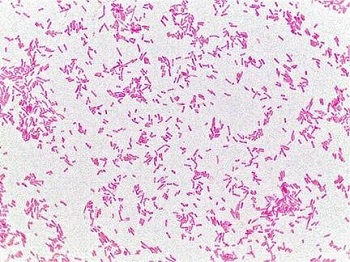
Fig: Gram-negative E. coli (Source: ResearchGate)
Result of Gram Staining
The organisms after gram staining which have retained deep violet color from crystal violet are Gram-positive microorganisms.
The organisms after gram staining that have lost crystal violet and taken safranin to appear red or pink in color are gram-negative microorganisms.