Laboratory Diagnosis of Fungal Infections - Culture, Serologic test, Germ Tube Test
Laboratory diagnosis of fungal infection begins with sample collection, transportation, and processing of samples. Demonstration of fungal particles such as hyphae, and spores in the specimen confirms the diagnosis.
Samples for Laboratory diagnosis of fungal infections
Clinical specimens collected for the diagnosis of mycosis include:
Respiratory tract secretions
Cerebral spinal fluid
Blood
Tissue
Body fluids
Bone marrow
Urine
Vaginal secretions
Skin
Hair
Nail
Collection and transport of samples for Laboratory diagnosis of fungal infections
After the collection of samples following the sterile technique, it is stored in a sterile container within 2 hours. In case of delay, the specimen must be stored at 4°C.
In the case of solid-state samples, they are processed by centrifugation of softening/liquidisation and spread onto the desired agar medium.
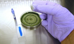
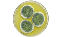
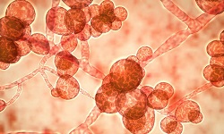
Fungal Culture
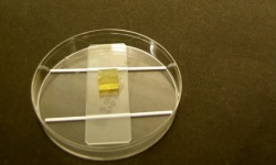
Medically, fungal culture is the method of growing yeasts and molds in the laboratory for research and/or diagnosis of fungal infections. For fungal isolation, cultivation, and maintenance, the most commonly used non-selective media is Sabouraud dextrose agar (SDA).
This method is a gold standard in mycology.
For the culture of fungi in a laboratory, the optimum temperature of the incubator should be maintained at 25 to 30°C.
In the case of dimorphic fungi, two sets of media must be inoculated and incubated. One set of media must be incubated at 37°C (for yeast form) and another set incubated at room temperature (25°C) for mold form.
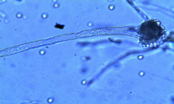
Some species of fungal colonies can be visualized within 24 to 72 hours after incubation. Such fungi include Candida, Aspergillus, Mucor, and Rhizopus.
On the other hand, many fungi can be slow-growing and may take up to 2-3 weeks to culture such as Histoplasma spp. Fastidious fungi should be incubated for 8 weeks.
Techniques for detection of culture:
Scotch Tape Preparation / Tape Touch Mount
Wet Mount Method for Fungi
Slide Culture for Fungi
Lactophenol Cotton Blue (LPCB) staining
Germ Tube Test
The germ tube test is a rapid, diagnostic test in which sample fungal spores are suspended in animal serum or egg albumin for three hours and microscopically examined for production of germ tubes. Other factors such as temperature, inoculum, strain, and medium also affect test tube production.
This screening test is mostly done to differentiate Candida albicans from other yeasts. Germ tube is one of the most important virulence factors for Candida albicans.
The germ tubes create somatic hyphae by the process of mitosis. This is done mostly when fungal culture produces colonies of white or cream color.
DNA Probes
Genetic probes such as DNA probes are commercially available for the detection of the presence of genetic materials for Blastomyces dermatidis, Coccidioides immitis, Cryptococcus neoformas, and Histoplasma capsulatum.
Serological test
The most commonly used serological tests for laboratory diagnosis of fungal infection include:
Immunodiffusion
Counter Immunoelectrophoresis (CIE)
Indirect fluorescent antibody (IFA)
Complement Fixation (CFT)
Radioimmunoassay (RIA)