Mitochondria - Introduction, History, Structure
Introduction to Mitochondria
Mitochondria is a type of cell organelle present in most eukaryotes such as fungi, plants, and animals. It is called the powerhouse of the cell as it uses aerobic respiration to generate adenosine triphosphate (ATP). The ATP is used throughout the cell as a source of chemical energy.
However, some cells lack mitochondria such as in mature mammalian red blood cells (RBCs). In unicellular organisms including microsporidia, parabasalids, and diplomonads, mitochondria have been reduced or transformed into other structures while members of the genus Monocercomonoides have lost their mitochondria.
.jpg)
Mitochondria (Source: Getty Images)
History of Mitochondria
The mitochondria were first discovered in 1857 by Swiss anatomist, histologist, and physiologist Albert von Kölliker. The organelle was found in the voluntary muscles of insects.
Decades later in 1898, the term mitochondrion was coined by microbiologist Carl Benda. It was later nicknamed “powerhouse of the cell” in 1957 by American cell biologist Philip Siekevitz.
Structure of Mitochondria
Mitochondria measures between 0.75 and 3 μm2 in cross-section and may vary in shape. It is double-layered – outer and inner membranes – and is composed of proteins and phospholipid bilayers. Both membranes have different properties and can be divided into five distinct parts.
Outer mitochondrial membrane
Intermembrane space
Inner mitochondrial membrane
Cristae
Matrix
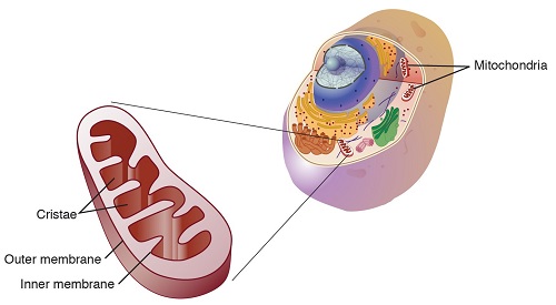
Mitochondria structure (Source: Genome.com)
Outer mitochondrial membrane
Encloses the mitochondria.
It is 60 to 75 angstroms (Å) in thickness.
Contains a large number of porins (integral membrane proteins).
Voltage-dependent anion channel (VDAC) is a primary transporter
Contains enzymes such as monoamine oxidase (MAO), kynurenine hydroxylase, fatty acid Co-A ligase, and rotenone-insensitive NADH-cytochrome c-reductase.
Intermembrane space
It is the mitochondrial intermembrane space between the outer membrane and the inner membrane.
Also known as perimitochondrial space.
This space is different from the protein composition of the cytosol since small molecules such as ions and sugars are permeable via the outer membrane but large proteins require specific signaling sequences to be transported across.
Inner mitochondrial membrane
Has a very high protein-to-phospholipid ratio.
Rich in inner membrane phospholipid, cardiolipin.
It contains proteins with three types of functions:
ATP synthase – generates ATP in matrix
Proteins responsible for electron transport chain redox reactions
Transport proteins that regulate metabolite passage to and from the mitochondrial matrix
Cristae
There are numerous folds that are compartmentalized, which expand the surface area of the inner mitochondrial membrane.
Increased surface area enhances the ability to produce ATP.
In typical liver mitochondria, the surface area is about five times as large as that of the outer membrane.
In cells with greater demand for ATP such as muscle cells, many cristae are present.
Folds are studded with oxysomes (F1 particles), and small round bodies.
Matrix
It is the space enclosed by the inner membrane.
Contains about 2/3 of the total proteins in a mitochondrion.
Has a high concentration of enzymes, tRNA, special mitochondrial ribosomes, and several copies of the mitochondrial DNA genome.
Important in ATP production with the action of ATP synthase present in the inner membrane.