Morphological classification of medically important fungi
Introduction to medically important fungi
Medically important fungi can be differentiated into numerous types.
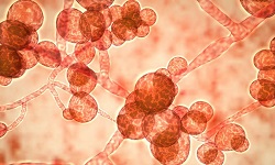
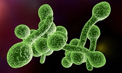
Yeast
Yeast are unicellular, oval, or round organisms whose size may range from 2 to 60 µm in diameter. Their colonies may be moist, creamy, opaque, or pasty on media. Some yeasts can be capsulated, which is a major virulence factor in pathogenic strains.
Most yeasts including Ascomycota, Basidiomycota, and Deuteromycota reproduce sexually and asexually. They produce blastoconidia (budding) and binary fission (mitosis) as asexual reproduction and sexually reproduce by production of ascospores and basidiospores.
Eg: Cryptococcus neoformans (pathogenic yeast)
Sacchaaromyces cerevisiae (non-pathogenic yeast)
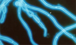
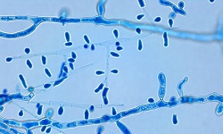
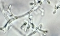
Yeast-like fungi
Yeast-like fungi grow partly as yeasts and partly as chains consisting of elongated budding cells joined end to end forming pseudohyphae. True hyphae consist of constriction at the septa. These septae are also present at the branching point.
Eg: Candida albicans
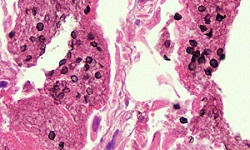
Dimorphic fungi
Depending on the growth conditions, dimorphic fungi can exist either as yeast or as filamentous molds. If dimorphism is temperature-dependent, it is called thermally dimorphic.
The yeast form i.e. parasitic phase occurs in host tissues with optimum growth temperature at 37°C while the filamentous form i.e. saprophytic form occurs in soil and grows at 22-25°C.
Systemic infections causing dimorphic fungi are as follows:
Histoplasma capsulatum (histoplasmosis)
Coccidioides immitis (coccidiomycosis)
Paracoccidioides brasilensis (paracoccidiodomycosis)
Blastomyces dermatitidis (blastomycosis)
Sporothrix schenckii (sporotrichosis)
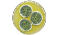
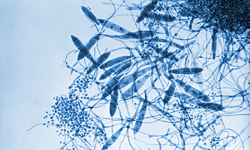
Molds
Molds are true mycelium that grow as branching filaments. They reproduce by producing both sexual spores and asexual spores. These spores may either develop from the vegetative mycelium or aerial mycelium.
Their hyphae are either septate or aseptate.
Septate fungi are morphologically coenocytic because the septa have holes through which the free flow of nuclei and other cytoplasmic material can occur.
Types of monomorphic molds on colony morphology:
White, cream, beige, or light ray surface, and black reverse
Eg: Chaetomium, Phoma, Trichophyton
White, cream, or light gray surface, non-pigmented
With maroconidia or microconidia
Eg: Fusarium spp.
Microsporum spp.
Trichophyton spp.
Verticillium spp.
Having only hyphae with chlamydoconidia
Eg: Microsprum spp.
Trichophyton spp.
Having arthroconidia
Eg: Coccidioides
Geotrichum spp.
Having sporangia
Eg: Mucor
Rhizopus
Absidia
Rhizopucor
White, cream, beige, or light gray surface, brown reverse
Eg: Trichophyton
White, cream, beige (light yellowish brown) or light grey surface, yellow, orange, or reddish reverse
Eg: Trichophyton
Microsporum
White, cream, beige, or light gray surface red to purple reverse
Eg: Penicillium
Trichophyton
Microsporum
Tan to brown surface
Having small conidia
Eg: Aspergillus
Batrytis
Cladosporium
Phialophora
Sparotrichum
Verticillium
Having large conidia or sporangia
Eg: Alternaria
Batrytis
Fusarium
Bipolaris
Yellow to orange surface
Eg: Aspergillus
Epidermophyton
Monilia
Penicillium
Sporotrichum
Trichophyton
Green, dark gray, or black surface, dark reverse
Having small conidia
Eg: Botrytis
Wangiella
Having large conidia
Eg: Alternaria
Bipolaris
Pithomyces
Having only hyphae (with or without chlamydoconidia)
Eg: Piedraia
Madurelia
Having large fruiting bodies
Eg: Chaetomium
Phome spp.
Green surface, light reverse
Eg: Gliocladium
Penicillium
Verticillium
Epidermophyton
Aspergillus
Dark grey or black surface, light reverse
Eg: Aspergilus
Syncephalastrum
Pink to violet surface
Eg: Acermonium spp.
Aspergillus spp.
Fusarium
Microsporum
Monilia