Mycobacterium leprae - Pathogenesis, Host immunity, Virulence Factors
Pathogenesis of Mycobacterium leprae
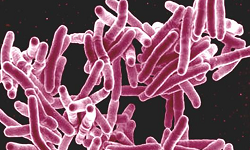
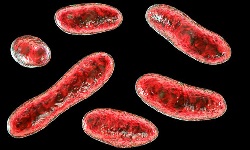
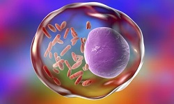
Mycobacterium leprae is an obligate intracellular bacillus with an affinity for macrophages and Schwann cells.
Mycobacterium leprae infects Schwann cells in nerves leading to demyelination (damage of myelin) sheath thereby damaging the nerve cells and loss of sensitivity. Involvement of mucosal cells leads to disfigurement and erythematous and macules appear on the skin due to loss of pigmentation.
Virulence factor of Mycobacterium leprae
Phenolic glycolipid – 1 (PGL-1) is a prominent surface lipid found on the outer layer of the cell wall and a major virulence factor. The lipid is specific to Mycobacterium leprae and is the best-studied virulence factor of Mycobacterium leprae. PGL-1 protects the lepra bacillus, once inside the phagocytic cells, from oxidative killing by macrophages by removing hydroxyl radicals and superoxide anions.
Pathogenesis and disease leading to tissue damage depend on the following factor:
CMI of the host
Multiplication and spread of lepra bacilli
Development of immunological complications such as lepra reaction leading to tissue damage
development of nerve damage
Mycobacterium leprae replicates intracellularly in the skin history and nerve cells (Schwann's cell) and has two forms- tuberculoid leprosy and lepromatous leprosy.
Tuberculoid leprosy induces a cell-mediated response that limits its growth. In this form, it multiplies at the site of entry, usually the skin, invading and colonizing Schwann cells.
Mycobacterium leprae can invade Schwann cells with a specific laminin-binding protein of 21 kDa in addition to PGL-1. Schwann cells are a major target for infection by Mycobacterium leprae to injury of the nerve, demyelination loss of axonal conductance, and consequently disability. High level of the T-cell immune response. The microbes then induce TH lymphocytes, epithelial cells, and giant cell infiltration of the stein causing infected individuals to exhibit large flattened patches with raised and elevated red edges on their skin. These patches have dry, pale, hairless centers, accompanied by a loss of sensation.
In lepromatous form, microbes' proliferation with the macrophages can be mediated by complement receptors CR1 (CD 35), CR (CD11b/CD 18), and CD4 (CD11C/CD18) and is regulated by protein kinase. It grows within the epithelial tissues of the face and ear lobes. CMI impaired large numbers of Mycobacterium leprae in the macrophages and infected patients develop papules at the entry site, marked by a folding of the skin.
Extensive penetration of these microbes may lead to severe body damage: for eg: loss of bones, fingers, and toes.
Host Immunity against Mycobacterium leprae
Cellular/Host immune responses aginst Mycobacterium leprae are poor/absent.
.jpg)
.jpg)