Mycoplasma hominis - Laboratory Diagnosis, Sample, Transport, Microscopy, Culture, Serodiagnosis
Laboratory diagnosis of Mycoplasma hominis
The laboratory diagnosis of Mycoplasma hominis is done after collection of samples.
Sample
Genital secretions/swabs
joint fluids
amniotic fluid
blood
semen
pleural secretions
urine
tissue, and wound aspirates
* For isolation of Mycoplasma hominis, the sample is pre-enriched in Mycoplasma broth (which contains arginine and urea). It also contains 10 microgram/ml of erythromycin which makes it selective for Mycoplasma hominis.
Transport
Since Mycoplasma hominis do not have a cell wall, they are highly susceptible to drying. Thus, transport media is required in cases of delayed examination. Suitable transport media such as SP4 transport medium can be used to prevent desiccation
If the storage time is expected to exceed 24 hours prior to cultivation, the sample + transport medium should be frozen at -80°C.
Microscopy
Mycoplasma hominis is cell-wall deficient so the gram stain is poorly observed in microscopy- hence not done
Acridine orange or fluorochrome stain can be used. But they are non-specific stains and will stain other bacteria as well as human cells with nucleic acid
Direct Ag-detection
Direct Ag-detection Mycoplasma hominis can be done by:
Direct/Indirect immunofluorescence
Counter-current immunoelectrophoresis
Immunoblotting
Culture
* Due to its fastidious nature and slow growth (3-4 weeks), culture is not generally done in routine diagnosis.
However, Mycoplasma hominis can be cultured in selective media to prevent the overgrowth of faster-growing organisms and is incorporated with Urea or arginine. If color change is observed (pH change), a 0.1-0.2ml aliquot is subcultured to fresh broth or agar media.
After enrichment, it is transferred in A8 agar and incubated in an atmosphere of 95% N2 and 5% CO2 at 35-37°C for 24-48 hours. Fried egg colonies are observed on A8 media which are 20-300 micrometers in diameter.
Media used for the culture of Mycoplasma hominis are => A7 and A8 agar medium; Triphasic system (Modified) NYC medium; Shepards 10B broth SP4 glucose broth with arginine or urea.
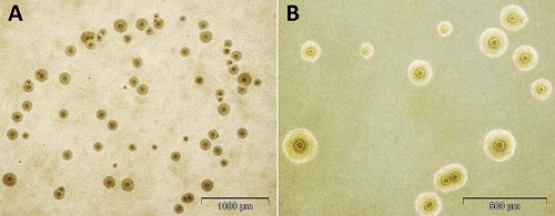
Fig: M. hominis colony (Source: Urology Gold Journal)
Identification of bacteria
The identification of Mycoplasma hominis is done by the following methods:
Agar is examined at 24-72 hrs following incubation
Mycoplasma hominis colonies are large (20-300 micrometers in diameter) and are urease negative with a fried-egg appearance
On BA, it produces non-hemolytic, pinpoints colonies that do not stain with gram’s stain but do stain with Dienes or acridine orange stains.
Serodiagnosis
The methods used for serodiagnosis of Mycoplasma hominis include:
CFT (Complement Fixation Test)
ELISA (enzyme-linked immunosorbent assay)
EIA (Enzyme immunoassay)
Indirect immunoflurescent Ab test
Indirect hemagglutination (rarely used)
Metabolism inhibition (rarely used)
Confirmatory test
The Mycoplasma hominis can be confirmed by the following test:
Growth inhibition by homologous antisera
Molecular diagnosis- PCR
AST, Treatment of Mycoplasma hominis
Mycoplasma hominis is resistant to erythromycin and occasionally to tetracyclines. In such cases of AST and treatment, clindamycin is used in treatment.