Mycoplasma pneumoniae - Lab diagnosis, Culture, Antigen, Diene, Hemadsorption, Tetrazolium reduction test
Laboratory diagnosis of Mycoplasma pneumoniae
A definitive laboratory diagnosis of Mycoplasma pneumoniae takes 3-4 weeks. Hence, treatment is started without waiting for lab results.
Specimen
Throat washing, bronchial washing, and expectorated sputum are common specimens/samples collected for the detection of Mycoplasma pneumoniae. Tracheal washings are more useful than sputum specimens as patients do not produce any sputum
Since mycoplasmas including Mycoplasma pneumoniae do not have a cell wall, they are highly susceptible to drying. Thus, transport media is required in cases of delayed examination. Suitable transport media such as SP4 transport medium can be used to prevent desiccation
If the storage time is expected to exceed 24 hours prior to cultivation, the sample + transport medium should be frozen at -80°C.
Microscopy
Mycoplasma pneumoniae is cell wall deficient. So they are stained poorly by gram’s stain and not visualized properly on microscopy. Acridine orange or a fluorochrome stain may be used but these non-specific stains will stain the nucleic acid of bacteria as well as human cells.
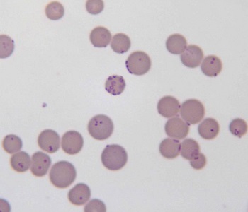
Fig: M. pneumoniae microscopy (Source: Wikipedia)
Direct antigen (Ag) detection
Mycoplasma pneumoniae infection can be diagnosed by direct antigen (Ag) detection
(In) Direct immunofluorescence
counter- current immunoelectrophoresis
immunoblotting: with monoclonal Ab are the tests used for the detection of Ag in clinical specimens
Culture
Mycoplasma pneumoniae culture is of little practical value because of its fastidious growth requirements and the need for 3-4 weeks for the culture. The medium must be selective to prevent the overgrowth of faster-growing organisms that may be present in a clinical sample. Some examples of media used are:
Biphasic SP4
Triphasic system
PLO broth or agar with yeast extract + horse serum
Modified NYC medium
Incubation is done at 37°C in presence of 95% N2, %5 CO2, and Mulberry-shaped homogenous colonies appear.
Different metabolic activity of Mycoplasma pneumoniae for the different substrates is used to detect their growth.
Eg: Glucose (Dextrose) is incorporated into the media. It ferments glucose to lactic acid and results in a change detected by a color change in a dye indicator. (Red => yellow colour; phenol red)
Identification of bacteria
On agar, Mycoplasma pneumoniae will appear as spherical grainy, yellowish forms that are embedded in the thin outer layer.
Color change
Different metabolic activity of Mycoplasma pneumoniae for the different substrates is used to detect their growth.
Eg: Glucose (Dextrose) is incorporated into the media. It ferments glucose to lactic acid and results in a change detected by a color change in a dye indicator.
(Red => yellow colour; phenol red)
Diene test
The procedure of the diene test is as follows:
I. Add Diene stain directly to a plate containing suspected colonies
ii. Plate immediately rinsed with d/w (stain removed)
iii. Add 1ml 95% ethanol and keep for 1 minute (decolorized)
iv. Plate re-washed with d/w and allowed to dry
v. colonies observed under the microscope
Fried-egg appearance colonies appear highly granular with the center of dark blue and periphery stained light blue agar that appears clear or slightly violet
Mycoplasma other than Mycoplasma pneumoniae becomes colorless after a certain time because it reduces the methylene blue
Inhibition of their growth with specific Mycoplasma pneumoniae antisera confirms the colonies.
Haemadsorption test
The steps followed for the haemadsorption test for Mycoplasma pneumoniae are listed below.
Mycoplasma pneumoniae colony on agar + 2 ml 0.2-0.4% guinea pig RBC
Incubated at 35°C for 35 mins
gently wash with 3ml of Mycoplasma growth medium
washing fluid gently aspirated by pipette
Mycoplasma pneumoniae adsorbs RBC on the surface which is observed under a microscope at 40%
Tetrazolium reduction test
This tetrazolium reduction test is based on the principle that Mycoplasma pneumoniae can reduce triphenyl tetrazolium to formazan.
Triphenyl tetrazolium (colorless compound) ========> formazan (red-colored compound)
Food colonies on agar with a solution of 2-p-iodophenyl 3-nitrophenyl-5- phenyltetrazolium chloride
Incubate at 35°C for 1 hour
Mycoplasma pneumoniae colonies appear reddish and may appear purple to black for 3-4 hours of incubation
Serodiagnosis
Serodiagnosis is based on the demonstration of specific Ab in serum using Mycoplasma pneumoniae antigen.
Complement fixation tests and ELISA tests are frequently used.
Complement fixation test
recent infection is detected by $ fold rise in complement-fixing Ab titer or a single titer of 1:64 or more
The complement-fixing Ab appears after 7-10 days of infection and reaches its peak after 4-6 weeks of infection
ELISA
IgM ELISA is used to detect specific IgM Ab in a serum specimen
with specificity = 99%, sensitivity = 97%
membrane-based ELISA is used for the demonstration of either IgM or IgG Ab. They are rapid (30 mins) and quantitative test
Indirect immunofluorescent Ab test
EIA
Non-specific serological test
Cross-reacting and non-specific Ag are used instead of specific Mycoplasma pneumoniae antigen (Ag). It includes:
Streptococcus MG agglutination test
Steps for the Streptococcus MG agglutination test include:
Heat-killed Streptococcus MG is used as Ag
This Ag is mixed with serial dilution of patients' unheated serum
incubation at 37°C overnight shows agglutination
Ab titer of 1:20 or more is suggestive of Mycoplasma pneumoniae infection
Cold agglutination test
human O group erythrocytes are used as Ag for cold agglutination test.
Based on the principle that autoantibodies that agglutinate human O group cells appear at low temperatures in most cases of atypical pneumonia
Molecular diagnosis
molecular diagnosis for the PCR assay detects various targets including 16rRNA sequence, insertion sequence, P1 adhesion, and organism-specific genes.
Specificity, fast turnaround time, and lack of need to cultivate fastidious organisms are particularly attractive assets.
It can also detect Mycoplasma pneumoniae at the early stages
AST, Therapy of Mycoplasma pneumoniae
The complex growth requirements of Mycoplasma pneumoniae have restricted their performances to a few labs.
Are susceptible to macrolides, tetracu=ycline, ketolides, fluoroquinolones, erythromycin
MDR mycoplasmas are seen in extragenital infections in immunocompromised patients
are resistant to β-lactams because cell walls absent
no vaccines