Negative Staining - Introduction, Principle, Purpose, Reagent, Procedure, Result
Introduction to Negative Staining
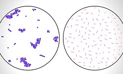
Negative Staining is a type of staining technique used to study the morphology and arrangement of bacterial cells which are difficult to stain spirillum/helical bacteria such as Campylobacter jejuni and Helicobacter pylori. It engages the use of acidic stains such as Indian ink or nigrosin and the results to yield a clear cell with a dark background.
Since heat fixation is not done, the cells are visible in their natural shape and size. Heat fixation is not required, and the cells are not subjected to the distorting effects of heat or chemicals.
Principle of Negative Staining
Negative Staining works due to the property of the stains/chromogens used. Both acidic chromogens and cell wall components, nucleic acids of bacteria are negatively charged. So due to the repulsion between the two negatively charged units, repulsion occurs and as a result, the dye will not penetrate the cell.
The unstained cells are easily visible against the colored dark background.
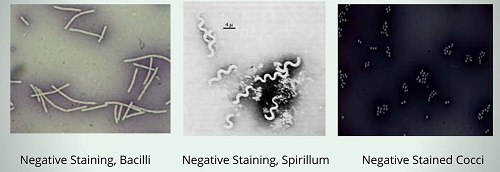
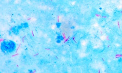
Fig: Negative Staining (Source: ResearchGate)
Purpose of Negative Staining
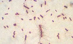
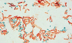
The negative staining method is an easy and quick method. It is used to study the morphology and arrangement of bacterial cells.
Reagent of Negative Staining
Any one of the following dyes/chromogens can be used as reagents for negative staining.
India ink
Nigrosin
Procedure of Negative Staining
The procedure of Negative Staining is as follows:
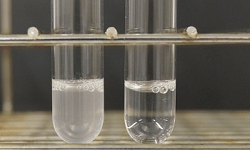
Place a drop of chromogen on one end of a clean grease free slide
Place one loopful of test organisms in the drop of the chromogen
Emulsify the mixture and make a thin smear with the help of another slide.
* Hold a second slide at 30° and place it on the mixture till the mixture spreads alongside the edge of the second slide touching it. Slide the second slide firmly, quickly, and in one single swipe over the first slide.
Let the smear air dry, do not use heat fixation or chemical fixation
Examine the slide under the microscope- first at low magnification and then at 100x under oil immersion.
Result of Negative Staining
The bacterial cells will be seen as clear/transparent with a blue background as a result of negative staining.

.jpg)
.jpg)
.jpg)