Nocardia spp. - Lab diagnosis, Prevention, Control, Treatment
Laboratory diagnosis of Nocardia
The laboratory diagnosis of Nocardia begins with the collection of specimens.
Sample
Sputum
respiratory secretion
skin biopsy
pus from abscess
Microscopy
Microscopy is done by ZN or Gram stain. Granules from pus collected are washed with saline, crushed between 2 slides, and stained by Giemsa or Z-N staining method.
Typical branching beaded filamentous hyphae of Nocardia are observed under the microscope.
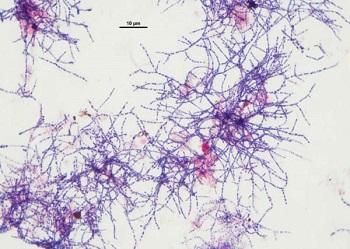
Fig: Nocardia spp. under microscope (Source: teamaquafix)
Culture
Nocardia spp. can be cultured on Brain Heart Infusion broth (BHI), Nutrient Agar (NA), Sabouraud Dextrose Agar (SDA), Thayer-Martin (with antibiotics). The inoculated medium is incubated at 36°C for 7-3 weeks.
Identification
Nocardia spp. can be identified by gram staining as well as ZN staining.
Biochemical tests are also used for the definitive identification of Nocardia spp.
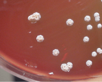
Nocardia spp. on BA (Image: ResearchGate)
Molecular
Molecular methods used for diagnosis of Nocardia includes
DNA probe
PCR
Prevention, Control, Treatment of Nocardia
The prevention, control, and treatment of Nocardia are listed below:
Prophylaxis with trimethoprim and sulfamethoxazole
Use of mask
xsulfaanamides, (sulfamethoxazole + teimethoprim), (imipenum + cilastation), (cefotaxime or ceftriazone + amikacin)