Rickettsia akari - Classification, Morphology, Clinical Manifestation, Culture
Classification of Rickettsia akari
Domain: Bacteria
Phylum: Pseudomonadota
Class: Alphaproteobacteria
Order: Rickettsiales
Family: Rickettsiaceae
Genus: Rickettsia
Species group: Spotted fever group
Species: akari
Clinical Manifestation of Rickettsia akari
Rickettsia akari is the causative agent of Rickettsial pox and is transmitted via mites.
The incubation period is 7 days. Symptoms include fever, myalgia, headache, and malaise. After 3-4 days of the appearance of fever, the generalized papular vesicular rash develops.
In 2-3 weeks, Rickettsia akari recovers with complete healing of the rash.
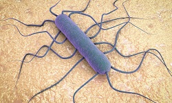
Morphology of Rickettsia akari
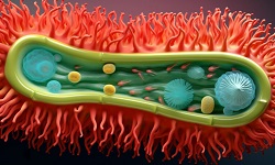
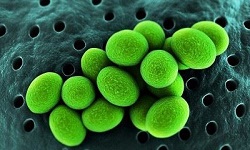
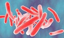
Morphologically, Rickettsia akari is gram-negative bacilli that are pleomorphic, obligate, intracellular, and fastidious with a size of 0.3 x 1-2 μm. It has a very small genome composed of 1-1.5 million bp. They multiply by binary fission within the cytoplasm of eukaryotic cells.
The bacteria are non-capsulated, non-motile, and contain a loose slime layer.
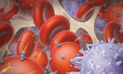
Rickettsia akari target cells in a host such as the reticuloendothelial system which releases mature rickettsiae resulting in the lysis of host cells. Rickettsiae are primary pathogens of arthropods (lice, fleas, ticks, mites) and are present in the intestinal tract- transmitted by arthropod vectors.
Rickettsia akari has LPS and peptidoglycan later. The LPS shows weak endotoxic activity. Moreover, they are poorly Gram-stained but stained well with Giemsa and Castaneda staining.
Culture of Rickettsia akari
Rickettsia akari fails to grow in cell-free media and grows in the cell line, chick embryo, and animal inoculation.
Cell lines such as HeLa, Hep 2, are used to maintain culture for primary isolation. Culture in yolk sack done for vaccine preparation. Animal inoculation is done in guinea pigs and mice. The optimum temperature 32-35°C,

.jpg)


.jpg)