Sabin-Feldman dye test - Introduction, History, Principle, Advantages, Disadvantages
Introduction of Sabin-Feldman dye test
Sabin-Feldman dye test is used to detect circulating antibodies (serologic test) in cases of Toxoplasma infection i.e. toxoplasmosis. It involves the evaluation of the development of new immunoassays in toxoplasmosis.
However, this test is used mostly in reference laboratories as a gold standard.
History of Sabin-Feldman dye test
Historically, in 1948, Sabin and Feldman described the first serological method, now known as the Sabin-Feldman dye test.
Principle of Sabin-Feldman dye test
The sabin-Feldman dye test is based on the presence of Toxoplasma antibodies and their detection in the sample. If Toxoplasma antibodies are present, the antibody-antigen complex activates complement to lyse the parasite membrane and the live tachyzoites present in this dye test are inactivated and killed.
As a result, the dead tachyzoites appear, thin, and distorted and do not retain color (colorless) in the presence of alkaline blue. This indicates a positive result for toxoplasmosis.
If Toxoplasma-specific antibodies are not present in the specimen, an antibody-antigen complex does not form and the tachyzoites with intact membranes are stained- appearing blue under a microscope. This indicates a negative result for toxoplasmosis.
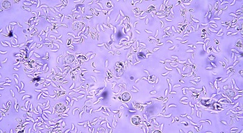
Image: The sabin-Feldman dye showing a positive result for Toxoplasmosis (Source: YouTube)
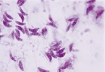
Image: The sabin-Feldman dye showing a negative result for Toxoplasmosis (Source: microbewiki)
Method of Sabin-Feldman dye test
The method/procedure of Method of Sabin-Feldman dye test include:
The sabin-Feldman dye test involves live tachyzoites, which are incubated with the test serum and accessory factor
* live tachyzoites are obtained from the peritoneal exudates of mice
* accessory factor, which is a complement in nature, is obtained from normal human serum
alcoholic solution of alkaline methylene blue (pH 11) is added to the mixture and re-incubated
the mixture is observed under a microscope for stained or unstained Toxoplasma tachyzoites
Antibody-titer of Sabin-Feldman dye test
The antibody titer of the test serum for of Sabin-Feldman dye test can be defined as the dilution of the serum at which half of the Toxoplasma tachyzoites (50%) are colorless (not stained/killed) while the other half of the Toxoplasma tachyzoites (50%) are stained (not killed).
As per the World Health Organization recommendation, antibody-titer is expressed in international units of ml of the serum (IU/ml).
Advantages of Sabin-Feldman dye test
The advantages of Sabin-Feldman dye test include:
The sabin-Feldman dye test has high sensitivity and specificity with no false positives reported so far.
If both Toxoplasma-specific IgM and IgA antibodies are detected in the serum, it indicates acute infection
* IgM antibodies are detected in the serum by IgM-IFA and double sandwich IgM-ELISA test.
IgM-IFA is able to detect IgM antibodies within the first few weeks of infection and if the antibody titer is 1:160 or more, it indicates acute infection.
the sandwich test has more sensitivity and specificity than the IgM-IFA method and if the IgM-ELISA has an antibody titer of 1:256 or more, it indicates acute infection.
Negative IgM test excludes recent infection
* presence of IgA antibodies in the test serum, aqueous or vitreous samples by the use of IgA-ELISA test also suggests an acute infection
Disadvantages of Sabin-Feldman dye test
The disadvantages of Sabin-Feldman dye test include:
live Toxoplasma tachyzoites are required
not a routine test but only used in reference laboratories
only detects Toxoplasma-specific IgG antibodies in the serum and since IgG antibodies can persist for years after infection, the test does not differentiate between recent or old infection
in immunocompromised individuals (AIDS, organ transplant patients) antibodies are produced in low levels and irregularly in the serum- and hence demonstration of IgG or IgM is not useful in such cases
cross-reactivity with Sarcocystis, Trypanosoma lewisi, and Trichomonas vaginalis has been reported