Schaeffer Fulton Staining - Introduction, Spore, Reagents, Principle, Procedure, Result
Introduction to Schaeffer Fulton Staining
Schaeffer Fulton Staining is performed to differentiate bacterial spores from other vegetative cells. It also functions to differentiate spore-forming bacteria from non-spore-forming bacteria.
Spore
Spores are highly resistant, metabolically inactive cell types that microorganism produces. Some spore-producing genera include anaerobes such as Clostridium, Desulfotomaculum, and aerobes such as Bacillus.
Usually, spores are formed when environmental conditions turn unfavorable to sustain a vegetative state such as scarcity in nutrients including carbon sources. These cells undergo sporogenesis, giving rise to an intracellular structure called an endospore, and are surrounded by impervious layers known as spore coats. As the environment continues to become unsuitable, endospores are released from the degenerating vegetative cells. Once released, these structures are called spores.
The chemical composition (calcium, dipicolinic acid) of spore layers makes it resistant to extreme desiccation, heat, freezing, radiation, and chemical components. When favorable environmental conditions return, these spores revert back to the metabolically active vegetative stage.
On the basis of the location of endospore, they are classified into three types:
central - spore located at the center of vegetative cell
subterminal - spore located between the center and edge of the microbial cell
terminal - spore located at the edge of the bacterial cell
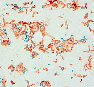
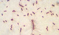
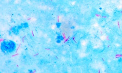
Fig: Microscopy after Schaeffer Fulton Staining (Source: ASM)
Reagents for Schaeffer Fulton Staining
Reagents used for Schaeffer Fulton Staining include:
Primary stain: Malachite green
Decolorizing agent: Distilled water
Counterstain: Safranin
Principle of Schaeffer Fulton Staining
The principle of Schaeffer Fulton Staining is based on the use of primary stain, decolorizing agent, and counterstain.
Primary stain
Malachite green is the primary stain of choice in Schaeffer Fulton staining. However, due to the impervious nature of the spore, malachite green cannot penetrate the microbial spores easily. Thus, heat is applied after the use of the primary stain and both vegetative cells and spores will appear green at this stage.
Decolorizing agent
When distilled water is used as a decolorizing agent, it cannot remove the primary stain inside the spore but easily washes out the stain in the cytoplasm of the microbial cells. Moreover, the malachite green does not have a strong affinity toward vegetative cell components. After this step, the vegetative cells will appear colorless.
Counterstain
The decolorized vegetative cells are now free to retain the counterstain i.e. safranin. The spores already have taken the primary stain and will not take the red color of the counterstain.
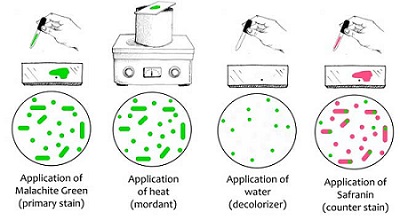
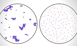
Fig: Schaeffer Fulton Staining procedure (Source: ASM)
Procedure
The procedure followed by Schaeffer Fulton Staining includes:
Take a clean, grease-free slide
Prepare a bacterial smear of the test organism
Allow the smear to air dry
Heat fix the dried smear by quickly passing the smear through the bunsen burner 2-3 times
Flood the smear with the primary stain (malachite green), place it in a water bath, and let it sit for 5 minutes. Add additional malachite green if the initial stain dries off
Let the glass slide cool and wash the smear with slow-running distilled water
Flood the smear with safranin (counterstain) for 20-30 seconds
Wash the smear with slow-running water
Gently tap the slide with blotting paper to remove the remaining water/dye
Examine under a microscope (oil immersion at 100x)
Heat fixation
Heat fixation is done in most staining procedures (except capsule staining, negative staining) because, during the washing steps, the microbial smear could wash away. During heat fixation, the bacterial proteins are coagulated and fixed to the glass slide surface.
It is done by rapidly passing the air-dried smear 2-3 times over a bunsen burner's flame.
* Instead of heat fixation, several methods of heat fixation have been recognized. One such commonly used procedure is the alcohol-based fixative. Ethanol or methanol can also be used as they also function by precipitating proteins.
Result of Schaeffer Fulton Staining
The spore-producing organism will be red colored with green colored endospore after Schaeffer Fulton Staining.
If the test organism is non-spore-producing, the entire cell will only be red in color.
.jpg)

.jpg)
.jpg)