Simple Staining - Introduction, Principle, Purpose, Reagents, Method, Result
Introduction to Simple Staining
Simple staining requires staining the bacterial cell directly with a positively charged dye/chromogens such as methylene blue, basic (carbol) fuchsin, and crystal violet which binds and stains the negatively charged surface of bacterial cells. Only a single reagent is used in simple staining.
Principle of Simple Staining
The principle of simple staining is based on positively changed chromogens are basic dyes. Since the nucleic acids and certain cell wall components of the microorganisms are negatively charged, it strongly attracts the cationic chromogen and binds with it.
If a negative stain is used, the bacteria remains unstained against a dark background because the negative stain repels the negatively charged surface of bacterial cells.
Purpose of Simple Staining
As simple staining is an easy and quick method, its principle is to observe the morphology and arrangement of bacterial cells.
Reagents of Simple Staining
Any one of the following dyes/chromogens can be used for Simple Staining.
Methylene blue
Carbol fuchsin
Crystal violet
Procedure of Simple Staining
The procedure of simple staining is as follows:
Take a clean, grease-free slide and prepare a bacterial smear of the test organism
Allow the smear to air dry
Heat fix the dried smear by quickly passing the smear through the bunsen burner 2-3 times
Flood the smear with any cationic chromogen depending upon the type of dye used
Carbol fuchsin = 15 to 30 seconds
Crystal violet = 20 to 60 seconds
Methylene blue = 1 to 2 minutes
Place the slide under slow-running water to wash off excess stain
Gently tap the slide with blotting paper to remove the remaining water/dye
Examine under a microscope (oil immersion at 100X)
Heat fixation
Heat fixation is done in most staining procedures (except capsule staining, and negative staining) because, during the washing steps, the microbial smear could wash away. During heat fixation, the bacterial proteins are coagulated and fixed to the glass slide surface.
It is done by rapidly passing the air-dried smear 2-3 times over a bunsen burner's flame.
* Instead of heat fixation, several methods of heat fixation have been recognized. One such commonly used procedure is the alcohol-based fixative. Ethanol or methanol can also be used as they also function by precipitating proteins.
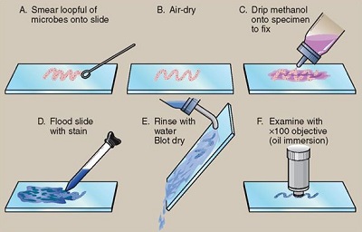
Fig: Simple staining (Source: Notes Hippo)
Result of Simple Staining
As a result of simple staining, the organism will be seen as red (Carbol fuchsin), violet (Crystal violet), or blue (Methylene blue).