Trichophyton mentagrophytes - Introduction, Classification, History, Habitat, Morphology, Culture, Identification
Introduction to Trichophyton mentagrophytes
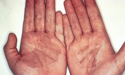
Trichophyton mentagrophytes are one of the most common fungi causing ringworm in pet animals and humans. It is the second most commonly isolated fungi causing tinea infections in humans. It is also one of the most common dermatophytes that cause zoonotic skin disease.
Trichophyton mentagrophytes can be isolated from cats, dogs, rabbits, and rodents (including guinea pigs). Human-to-human transmission occurs by some genetic variants e.g. Type VII and Type VIII. Distinct geographical ranges have particular genetic variants.
Classification of Trichophyton mentagrophytes
Trichophyton mentagrophytes is classified as follows:
Kingdom: Fungi
Division: Ascomycota
Class: Eurotiomycetes
Order: Onygenales
Family: Arthrodermataceae
Genus: Trichophyton
Species: T. mentagrophytes
History of Trichophyton mentagrophytes
Historically, Trichophyton mentagrophytes was first described by Robin Blanchard in 1853.
Habitat of Trichophyton mentagrophytes
Trichophyton mentagrophytes is a zoonotic disease transmitted to humans from companion animals (cats, dogs, rabbits, and guinea pigs). Although human-to-human transmission is uncommon, certain genetic variants e.g. Type VII and Type VIII are able to pass from infected human to another human.
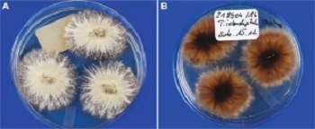
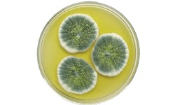
Trichophyton mentagrophytes on SDA - front view (left), reverse side (right) (Source: ResearchGate)
Morphology of Trichophyton mentagrophytes
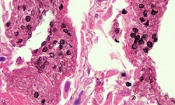
In non-living keratinized tissue, Trichophyton mentagrophytes form only hyphae and arthroconidia i.e. arthrospores. These arthrospores are individual cells fragmented from the hyphae.
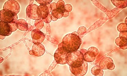
Trichophyton mentagrophytes colonies may range from granular to powdery. They produce abundant grapelike clusters of subspherical microconidia on terminal branches. In 30% of isolates, the hyphae are spiral while the thin-walled microconidia are smooth, club-shaped, and multiseptate. Each microconidium may measure 6 to 8 µm in diameter and 20 to 50 µm in length.
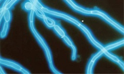
Macroconidia are thin-walled, smooth, club-shaped, and multiseptate. Depending on the strain of Trichophyton mentagrophytes, the number of macroconidia may be numerous or rare.
Culture of Trichophyton mentagrophytes
Like most fungi, Trichophyton mentagrophytes can be cultured in a laboratory.
It grows on Sabouraud’s Dextrose Agar (SDA) incorporated with chloramphenicol and cycloheximide and incubated at 25 to 30°C aerobically for 1 to 3 weeks.
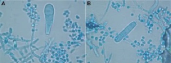
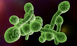
Trichophyton mentagrophytes macroconidia (Source: ResearchGate)
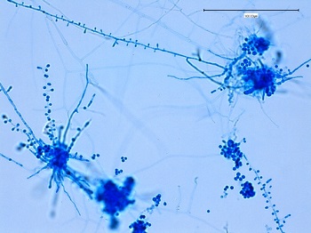
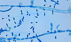
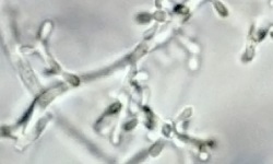
Trichophyton mentagrophytes microconidia (Source: Fun with Microbiology)
Identification of Trichophyton mentagrophytes
The fungi Trichophyton mentagrophytes are identified on the basis of colony morphology, color, pigment production, and presence/absence of microconidia or macroconidia.
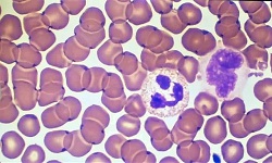
The fresh culture of fungi was extracted from the agar plate using cellophane tape and placed on a slide containing a drop of Lacto Phenol Cotton Blue (LPCB). The preparation is viewed under a microscope to demonstrate the presence/absence of microconidia or macroconidia.
Trichophyton mentagrophytes colonies may range from wooly, and granular to powdery. Reverse side colonies grown on SDA may be brown, yellow, or dark red in color while dark red color is absent in colonies grown in PDA containing !% glucose.
In 30% of isolates, the hyphae are spiral.
They produce abundant grapelike clusters of subspherical microconidia on terminal branches. The thin-walled microconidia are smooth, club-shaped, and multiseptate. Each microconidium may measure 6 to 8 µm in diameter and 20 to 50 µm in length.
Macroconidia are thin-walled, smooth, club-shaped, and multiseptate. Depending on the strain of Trichophyton mentagrophytes, the number of macroconidia may be numerous or rare.
Bromocresol purple (BCP) milk solid glucose agar test can also be used to identify Trichophyton mentagrophytes. Wood's lamp (blacklight) examination shows bright green to yellow-green fluorescence of hairs.