Corynebacterium diphtheriae - Laboratory Diagnosis
Lab Diagnosis of Corynebacterium diphtheriae
Initial treatment of Corynebacterium diphtheriae is based on clinical symptoms and treatment is started before the lab report. Hence, lab diagnosis is carried out for epidemiological purposes than for treatment.
Specimen
Specimen collected for diagnosis of Corynebacterium diphtheriae
Swab(s) from the nose, throat, biopsy tissue, etc.
pieces of pseudomembrane
* At least two swabs are collected- one for direct smear and the other for culture
Microscopy
Microscopy after gram staining reveals gram-positive bacilli in short chains
Albert, Neisser, or Ponder stain of direct smear shows metachromatic granules
Microscopy is not specific because other species of Corynebacterium are present as commensal in the throat
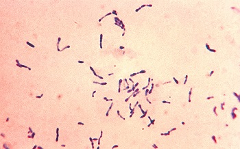
Fig: Corynebacterium diphtheriae (Source: CDC)
Culture
The culture of Corynebacterium diphtheria can be done in
Non-selective media: BA
Selective media: Tellurite blood agar
Enriched media: Loeffler medium, Tinsdale medium
* In Tellurite blood agar, reduces tellurite and produces grey or grey-black colonies measuring 0.5-2mm in diameter after 24-48 hours of incubation
* In tinsdale medium colonies are grey-black, raised, and surrounded by a dark brown area after 24-48 hours of incubation. The brown color is due to H2S production from the cystine interacting with tellurite. Commercial diphtheroids and other RT commensals colonies lack surrounding brown halo.
Biochemical test
Some biochemical tests performed for Corynebacterium diphtheriae are
Catalase => positive
Oxidase => negative
Nitrate test => positive
ferments glucose and maltose with acid production
few strains gravis and mitis biovars ferment sucrose
Corynebacterium diphtheriae gravis ferments starch with acid production
Identification of bacteria
3 Corynebacterium diphtheriae biotypes: mitis, intermedius, gravis
potentially toxigenic spp, have systinase but no pyrazinamidase activity
Toxigenicity testing
The following toxigenicity tests can demonstrate the production of toxins by Corynebacterium diphtheriae.
In vivo test:
subcutaneous test
Intradermal test
Subcutaneous test:
growth from an overnight culture of Corynebacterium diphtheriae on Loeffler’s slope is emulsified in 2-4 ml broth
Two guinea pigs are injected subcutaneously with 0.8ml of the emulsion. One of the guinea pigs is protected with 500 µl of diphtheria antitoxin injected intraperitoneally 18-24 hours before the test. Another guinea pig is not protected
If the test stain is virulent, the unprotected animal will die within 4 days. The protected guinea pig shall remain normal
Intradermal test:
0.1 ml of the emulsion(obtained from 18 hours of growth of test bacteria cultured on Loeffler's medium slope) is injected intradermally into shaven sites on each 2 guinea pigs
The control animal is given 500 µl of antitoxin the previous day. Another animal is given 50 µl of antitoxin intraperitoneally 4 hours after the skin test to prevent death
Toxigenically indicated by an inflammation reaction at the injection site progressing to necrosis in 48-72 hours in test animals. No change in the control of animal
In vitro test:
Elek’s gel ppt test
Tissue culture test
Elek’s gel ppt test:
- It is an immuno-precipitation test for demonstration of the biological activity of the toxin.
Procedure:
Using sterile forceps, soak a strip of filter paper in diphtheria antitoxin diluted to 1000 units per ml
Allow the strip to drain
Lay the strip in a petri dish. Leave the petri dish in an incubator for drying or for about 20 minutes
Prepare serum culture medium by adding 3 ml of clear sterile serum to 15 ml sterile cooled (50-55°C) protease peptone agar or Columbia agar.
Pour the serum agar medium into a petri dish containing an antitoxin strip and allow the medium to set firmly
Dry the medium at 35-37°C for 20-30 minutes and should not exceed 60 minutes.
Heavily inoculate test and control microorganism right angle to antitoxin strip and incubate at 35-37°C overnight
Precipitation lines are seen when viewed through a low-powered hand-magnifying lens
Tissue culture test:
Eukaryotic cell lines eg: African green monkey kidney, and Chinese hamster ovary are sensitive to diphtheria toxin
Inocular of test microorganisms in cell culture monolayer
The toxin produced by microorganisms diffuses and kills cells in the monolayer
Molecular test
PCR detects toxigenic strains of Corynebacterium diphtheriae from the clinical specimen
detects diphtherial toxin gene (TOX).
Sckick’s test:
The procedure for Sckick's test for Corynebacterium diphtheriae is as follows:
Inject 0.1 ml of highly purified toxin into 1 forearm
Inject 0.1ml of heat-inactivated toxin into another forearm as a control
Four types of reaction are observed:
* Positive reaction
* Negative reaction
* Pseudoimmune reaction
* Combined reaction
Positive reaction
the local inflammatory reaction that reaches maximum intensity in 4-7 days in the arm and reduces gradually
indicates the absence of an immunity to Corynebacterium diphtheriae
Negative reaction
Absence of any inflammatory reaction
indicates the presence of diphtheria antitoxin
individual immune to Corynebacterium diphtheriae infection
Pseudoimmune reaction
although the immune, allergic reaction is observed in both the test and control arm
inflammation reaches a peak in 36 hours and subsides in 72 hours in both arms
both immune and hypersensitive
Combine reaction
develops inflammation in the rest arm, which increases the intensity in 4-7 days
In the control arm, inflammation is seen maximum at 48-72 hours and subside
the person is not immune and hypersensitive