Coxiella burnetii - Classification, Introduction, Morphology, Epidemiology, Culture
Classification of Coxiella burnetii
Phenotypically, Coxiella burnetii can be classified as
Domain: Bacteria
Phylum: Pseudomonadota
Class: Gammaproteobacteria
Order: Legionellales
Family: Coxiellaceae
Genus: Coxiella
Species: burnetii
Introduction to Coxiella burnetii
Coxiella burnetii is poorly Gram-stained so it is stained with Giemsa stain. They multiply intracellularly in eukaryotic cells (monocytes and macrophages) and are transmitted by ingestion/inspiration of contaminated droplets.
Coxiella burnetii causes Q fever, an acute system infection primarily affects the lungs. It can survive extracellularly but can only grow in lung cells. It exists in 2 antigenic stages:
Phase I (large cell variant form)- isolated from animals and is highly infectious; metabolically inactive
Phase II (small cell variant form)- grown in cultured cell lines and are not infectious but acts like a spore assisting in the extracellular survival of the organism.
Also metabolically active
Q fever is a zoonotic disease transmitted from animals to humans. The smaller and more resistant to various chemical and physical agents than Rickettsia spp.
Morphology of Coxiella burnetii
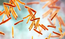
Coxiella burnetii is an obligate intracellular bacterium that is pleomorphic and 1x0.3 μm in size. Morphologically, they are gram-negative but stain poorly with Gram staining but stain best with Giemsa or Gimenez stain.
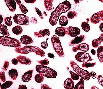
Fig: Coxiella Burnetii microscopy (Source: ResearchGate)
Reservoir, Source of Coxiella burnetii
Reservoirs for Coxiella burnetii include cattle, sheep, and goats. The organisms are shed in urine, feces, milk, and birth products. Humans are infected by the inhalation of contaminated aerosols and are resistant to desiccation and sunlight by forming spores.
Culture of Coxiella burnetii
In case of a laboratory-acquired infection, the Coxiella burnetii culture must be done in biosafety level 3 containment. However, the use of shell vial assay with human lung fibroblasts to isolate organisms from biopsy and buffy coat specimens has not resulted in lab-acquired infections.
Cultures are incubated for 6-14 days at 37°C in CO2 enriched environment and detected by using a direct immunofluorescent assay.
Related Article

Rickettsia rickettsii
Rickettsia rickettsii is a gram-negative, obligate intracellular bacillus t...
Rickettsia prowazekii
Rickettsia prowazekii is the causative agent of epidemic/louse-borne typhus...