Listeria monocytogenes - Lab diagnosis, Microscopy, Culture, Biochemical tests, Serology
Lab diagnosis of Listeria monocytogenes
Listeria monocytogenes infection can be diagnosed in the laboratory with a collection of samples.
Sample
CSF
Blood
leftover food
* (majority of tests will be carried out on food and environmental samples)
* samples should be processed asap. If not possible store at 4°C
Microscopy
The microscopy of Listeria monocytogenes shows gram-positive, rods in chains that are non-capsulated, and non-sporing.
The motility test shows tumbling motility at 22-25°C but immobile at 37°C.
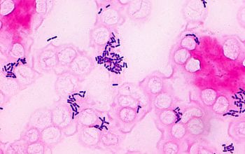
Fig: Listeria monocytogenes gram-stain (Source: Microbe Canvas)
Culture
The culture of Listeria monocytogenes is based on the origin of the samples/specimens.
Clinical samples: they are directly cultured onto selective media-modified oxford agar or BA. Incubation is done at 37°C for 24-48 hours.
Environmental/food samples: First inoculated into a pre-enrichment culture medium such as Buffered Listeria Enrichment Broth (BLEB) or Half-Fraser broth. The incubation is done at 30°C for 24 hours.
Colony appearance: If grown in BA, it shows β-hemolysis
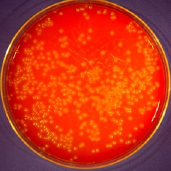
Listeria monocytogenes on BA
Biochemical tests
Some biochemical tests of Listeria monocytogenes include:
Catalase +ve
Oxidase -ve
Esculin hydrolysis test +ve
ferments/use glucose, maltose, salicin with acid production but no gas
CAMP +ve and enhanced in the vicinity of S. aureus
Serology
Some serological tests used for Listeria monocytogenes lab diagnosis are
EIA
ELISA-based assay incorporating fluorescent colorimeter detection
Molecular methods
A few molecular methods used for the diagnosis of Listeria monocytogenes are as follows:
DNA hybridization
real-time PCR