Mycobacterium leprae - Classification, Morphology, Culture, Cell wall, Antigen, Characteristics
Classification of Mycobacterium leprae
Mycobacterium leprae can be classified phenotypically by:
Domain: Bacteria
Phylum: Actinomycetota
Class: Actinomycetia
Order: Mycobacteriales
Family: Mycobacteriaceae
Genus: Mycobacterium
Species: lepra
Characteristics of
Mycobacterium leprae causes leprosy, a chronic infectious disease that affects the skin, peripheral nerves, and mucosa of the upper respiratory and the eyes. It is the only mycobacterium known to cause infection of the nervous system. It is mainly transmitted via the respiratory tract or skin and has a long incubation period (2-5 years).
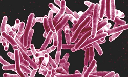
Morphology
Mycobacterium leprae is less acid-fast than Mycobacterium tuberculosis. Hence, 5% sulphuric acid, instead of 20% is used for decolorization. The bacterium is non-motile, non-sporing, gram-positive, and stains more readily than Mycobacterium tuberculosis.
Morphologically, Mycobacterium leprae also exhibits cubical, lateral, and even branching forms measuring 1-8 x 0.2-0.5 μm. It is a slightly curved or straight rod with parallel sides and surrounded ends. It is an obligate intracellular bacterium that multiplies in tissues at a cooler temperature.
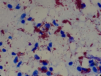
Fig: Mycobacterium leprae microscopy (Source: Wikipedia)
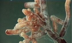
Cultural characteristics of Mycobacterium leprae
Mycobacterium leprae has not been cultured in-vitro like bacteriological media and tissue culture
Footpads of mice
footpads of mice are used as a standard procedure for experimental works of Mycobacterium leprae
intradermal inoculation of lepra bacilli into the footpads of mice results in the development of granuloma at the site of inoculation in 1-6 months
Nine-banded armadillo
the nine-banded armadillo is highly susceptible to Mycobacterium leprae
Mycobacterium leprae in these animals produces a lesion typical of lepromatous leprosy
This animal is now used for genetic studies including the development of vaccines
Cell wall components of Mycobacterium leprae
The cell wall components of Mycobacterium leprae stimulate the production of antibodies and the cell-mediated response. The cell wall of Mycobacterium leprae consists of four layers:
Peptidoglycan is the innermost layer, which gives the bacteria rigidity and shape
Liparabinomannose-B (LAM-B) is the latter next to the peptidoglycan layer
Mycolic acid is the third layer attached to the LAM-B membrane
Mycosides are the outermost fourth layer of the cell wall. Phenolic glycolipid (PGL-1) is the major component of the outermost layer of the cell wall.
Antigenic structure of Mycobacterium leprae
Mycobacterium leprae contains a wide variety of mycobacterial Ag. LAM-B is a major Ag of Mycobacterium leprae and is highly immunogenic and induces a high level of serum antibodies. PGL-1 is also the major antigen component of the outermost layer of the cell wall.

.jpg)
.jpg)