Bacteroides fragilis - Lab Diagnosis
Lab Diagnosis of Bacteroides fragilis
Anaerobes such as Bacteroides fragilis form part of normal flora. Therefore, the presence of an anaerobe in the sample does not indicate its role in disease as facultative anaerobes die on exposure to O2, proper care should be exercised to minimize the contact with air during collection, transport, and handling of the specimen.
The laboratory diagnosis of Bacteroides fragilis includes:
Sample
Blood
CSF
Bone marrow
joint fluids
material aspirated from abscesses
BW obtained with a double-lumen plugged catheter, pleural fluid, direct lung aspirate
Sample collection and transport
The specimen collected for Bacteroides fragilis diagnosis may be collected by aspiration of the specimen into an airtight syringe. After collection of the specimen, the needle is plunged into a sterile rubber cork (seal) and immediately sent to the lab.
An anaerobic transport system for liquid specimen/swab specimen for Bacteroides fragilis diagnosis tissue specimen can be used. In an anaerobic transport medium for liquid specimens, the specimen is injected into the tube through the rubber septum containing an agar indicator system.
Anaerobic transport medium for swab specimens contains a sterile swab and O2 free inner tube. After the sample has been collected for Bacteroides fragilis diagnosis, the swab is inserted back into the inner tube with an agar indicator system on the bottom of the outer tube.
For tissue specimens, tissue is placed in a small amount of saline (to keep it moist). GasPack Pouch can be used for purpose of transporting tissue.
Macroscopic examination
Specimens for Bacteroides fragilis inspection should be inspected for characteristics that indicate the presence of anaerobes (foul odor, sulfur granules, brick red fluorescence under UV light).
The foul odor is not present in the case of Bacteroides fragilis.
Gram staining
Bacteroides fragilis are gram-negative, pleomorphic rods with rounded ends, occur singly or in pairs
Gram-negative anaerobes stain poorly with safranin. Thus safranin (counterstain) must be left for 3-5 minutes. Alternately, 0.5% aq. Basic fuschin can be used as a counterstain
Gram enhancer, applied after the decolorization step, suppresses the red color in the background, aiding the differentiation of gram-negative anaerobes.
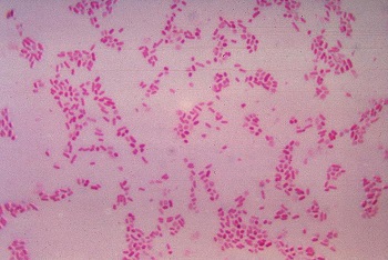
Fig: Bacteroides fragilis gram staining (Source: Wikipedia)
Culture
The culture of the specimen is performed in solid media and liquid media such as Ana BAP (Anaerobic blood Agar Plate) containing 10-20 µg gentamycin, Bacteroides Bile Esculin (BBE), Lacked kanamycin Vancomycin (LKV) can be used to isolate Bacteroides fragilis (selective LKV).
Two types of liquid medium are used for the isolation of Bacteroides fragilis - Robertson’s cooked meat medium and Thioglycolate medium.
In addition, BA, CA, and MA are also used because most anaerobic infections are polymicrobial and may include aerobic and facultatively anaerobic bacteria.
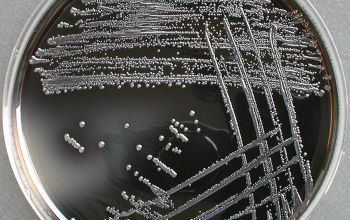
Fig: Bacteroides fragilis colonies on BBE (Source: ResearchGate)
Incubation condition and duration
Inoculated plates were immediately incubated under anaerobic conditions at 35-37°C for 48 hours
Incubation for at least 5 days should be done before discarding
Plates may be removed from the anaerobic environment for a brief evaluation
Thioglycollate broth can be incubated anaerobically with the cap loose or cap tight. The broth should be inspected daily for 7 days.
* (should not be exposed to O2 until after 48 hours of incubation as anaerobes are most sensitive to oxygen during the log phase)
Observation of colonies and presumptive identification
On AnaBAP: white to gray, circular, entire, convex, translucent to semiopaque, non-hemolytic
On BBE: colonies are >1 mm, circular, entire, raised, and either
low convex, dark gray, friable, surrounded by a dark gray zone (esculin hydrolysis) and sometimes a precipitate (bile)
glistering, convex, light to dark gray, and surrounded by a gray zone
The growth on liquid medium (Thioglycollate broth / Robertson cooked meat medium) is indicated by a change in turbidity
Subculture is done from these mediums in Ana BAB, BBE, and further processing is done.
Confirmation of Bacteroides fragilis
Confirmation of Bacteroides fragilis is done by following methods:
Gram stain: gram-negative rods reveal Bacteroides fragilis
Biochemical tests:
Sucrose positive
Glucose positive
Lactose positive
Maltose positive
growth in 20% bile
Eschulin hydrolysis positive
Catalase positive
Indole negative
Oxidase negative