Brugia timori - Clinical Manifestation
Clinical Manifestation of Brugia timori
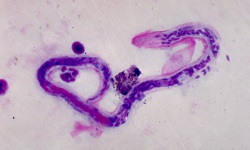
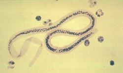
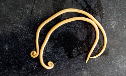
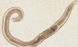
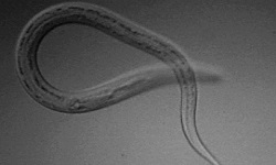
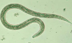
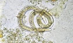
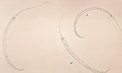
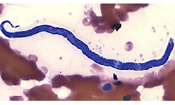
Brugia timori is the causative agent of disease Timor filariasis or Timorian filariasis. In humans, Brugia timori may manifest as:
Endemic normal
Asymptomatic stage
Acute stage
Chronic stage
Endemic normal
In areas endemic to filariasis caused by Brugia timori, a portion of the population does not show any overt clinical manifestation of the disease. Such a population may also lack microfilariae in the blood even if they are exposed to the infective third-stage larvae (L3).
However, it is difficult to demonstrate if the said population is uninfected by Brugia timori or simply has undetected filarial nematode infection.
Asymptomatic stage
Individuals in the asymptomatic stage of Brugia timori infection have microfilariae in their blood but lack any clinical manifestations of filariasis. Such individuals may remain asymptomatic for years and even for a lifetime. If such individuals settle in non-endemic filarial areas, they may spontaneously become microfilaraemia.
The asymptomatic stage in individuals has been suggested to have been caused by the downregulation of the TH1 inflammatory component of the immune response while the TH2 is stimulatory. It also involves depressed cytokine IFN-c but elevated IL-4 levels (IL4 suppresses activation of TH1).
After several years, hyporesponsiveness breaks down and the appearance of inflammatory reactions begins.
Acute filariasis
The inflammatory phase i.e. acute filariasis is caused by antigens released from the female Brugia timori parasite while microfilariae are not responsible for inflammatory changes.
This condition manifests as:
Filarial fever
Lymphadenitis
Lymphoedema
Filarial fever
filarial fever due to Brugia timori infection is usually low grade
sometimes filarial fever may be accompanied by chills and become severe
symptoms include general malaise, pain, headache
episodes of filarial fever may occur several times a year – which lasts upto a week each time
Lymphoedema
lymphoedema is caused by the presence of adult worms in the lymphatic channels
lymph flow is hindered by the Brugia timori parasite
individuals with lymphoedema suffer from periodic attacks of filarial fever, adenolymphangitis, and lymphadenitis
Lymphadenitis
in lymphadenitis, the lymph nodes are inflamed
commonly infected lymph nodes include epitrochlear, cervical, axillary, inguinal, abdominal, pelvic, supraclavicular
unusual lymph nodes infected include wrist, iliac, pectoral, creat, mediastinal, intercostal, mid humoral
infected lymph nodes are tender, enlarged, and matted but are not attached to the skin
such lymph nodes may or may not containBrugia timori parasites
lymph nodes capsules are thickened with fibrotic septa while the middle layer of the node may contain dead or living male and female adult Brugia timori filarial nematode
scar tissue is present in the outer layers
Chronic filariasis (Obstructive phase)
Chronic filariasis, also known as the obstructive phase, takes 10 to 15 years to develop after an individual is infected with Brugia timori. During this stage, acute inflammation subsides while fibrosis advances as the parasitic worms die, are absorbed, or are calcified.
Microfilariae are usually absent in chronic cases but filarial abscesses are common. Clinical manifestations of chronic filariasis caused by Brugia timori include elephantiasis.
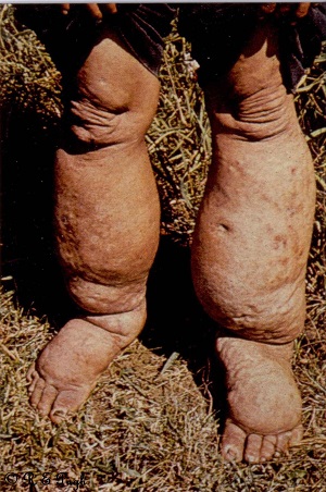
Brugia timori elephantiasis (Source: parasite.org.au)
Elephantiasis
elephantiasis is a characteristic feature of chronic Brugia timori infection
caused by complex immune reactions for a long duration as well as repeated infections over many years
this manifestation is seen only in a small percentage of infected individuals in endemic areas
in males, elephantiasis occurs in the scrotum, legs, and arms
in females, elephantiasis is seen in arms and legs
if legs and arms are infected, swelling is seen below the knee and below the elbow respectively
at first, swelling is pitting which later becomes non-pitting
the elephantoid mass consists of fibrous tissues containing fat
overlaying skin of the leg, the scrotum becomes thick, warty, and fissured
ulceration and secondary infection with fungi or bacteria also may occur
peripheral blood usually lacks Brugia timori microfilariae
.jpg)