Dracunculus medinensis - Introduction, Classification, History, Habitat, Morphology
Introduction to Dracunculus medinensis
Dracunculus medinensis is also known as serpent worm, guinea worm, or medina worm. It is the etiological agent of dracunculiasis or dracontiasis and is characterized by a chronic cutaneous ulcer on the part of the body that frequently comes in contact with water.
Classification of Dracunculus medinensis
The classification of Dracunculus medinensis is as follows:
Kingdom: Animalia
Phylum: Nematoda
Class: Secernentea
Order: Camallanida
Family: Dracunculidae
Genus: Dracunculus
Species: D. medinensis
History of Dracunculus medinensis
In 1853, Baston first described the morphological feature of the spirurid nematode. Russian biologist Fedtschenko discovered cyclops as vectors causing transmission of the disease.
Habitat of Dracunculus medinensis
Adult Dracunculus medinensis females inhabit the subcutaneous tissues of humans’ feet or lower limbs. The parasite may also inhabit other parts of the body such as the head and neck.
Morphology of Dracunculus medinensis
The morphological features of Dracunculus medinensis include adult worm, first-stage larva, and infective form.
Adult form
The adult form of Dracunculus medinensis includes separate male and female parasites.
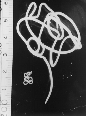
adult Dracunculus medinensis male (left) and female (right) - scale in cm (Source: ResearchGate)
Male
adult male of Dracunculus medinensis die immediately after fertilizing female
are difficult to demonstrate in the specimen
measures 15 to 40 mm in length and 0.44 mm in diameter
are obtained from experimental animal infections
life-span is around three to six weeks
Female
female Dracunculus medinensis is one of the longest nematodes infecting humans
measures 50 to 120 cm in length and 0.7 to 1.7 mm in diameter
milky white, slender, resembles a thick twine of thread
rounded anterior end, tapering pointed posterior end forms a hook-like structure
the anterior end has a minute triangular mouth
mouth surrounded by an inner layer of 4 to 6 papillae and an outer layer of 4 pairs of papillae
the female genital tract consists of a pair of uteri, tubules, oviducts, and a single unpaired vagina
viviparous
uterus completely filled with thousands of eggs, embryos, and first-stage larvae
over a period of 3 weeks, larvae laid in successive batches through the vaginal opening
lives for about a year – longer life-span than males
First-stage larva
first stage larva of Dracunculus medinensis is unsheathed, coiled with a round anterior end long slender filariform tail
large in size
measures 650 to 750 mm in length and 17 to 20 mm in diameter
conspicuous striated cuticula
locomotion takes place with a stiff motion, briskly coiling and uncoiling body – shows tadpole-like movement
the short life span of 6 days – dies if not taken up by cyclops
if taken up by cyclops, further development of Dracunculus medinensis takes place in cyclops
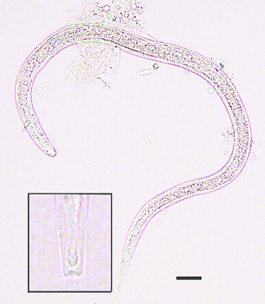
third-stage Dracunculus medinensis larvae (Source: ResearchGate)
Infective form
The infective form of Dracunculus medinensis is the third-stage larva. It is found in the body cavity of cyclops.