Dracunculus medinensis - Life Cycle, Pathogenesis, Pathology, Host Immunity, Clinical Manifestations
Life Cycle of Dracunculus medinensis
The life cycle of Dracunculus medinensis is completed in two classes of the host, definitive host, and intermediate host.
Definitive host: Humans
Intermediate host: Cyclops (twelve species including Mesocyclops leuckarti, Mesocyclops hyalinus)
humans acquire Dracunculus medinensis infection after drinking unfiltered water infested with cyclops
third-stage Dracunculus medinensis larvae are released in the host stomach, which then migrates to the small intestine
in the small intestine, the parasitic larvae penetrate the small intestinal mucosa to reach retroperitoneal tissue
in the human host tissue, the parasite molts develops, and sexually matures into adult male and female within 6 months
male Dracunculus medinensis parasite dies after fertilizing the female in the subcutaneous tissue
following fertilization, the gravid female migrates deep into the host tissue of the parts of the body which usually come in contact with water
female remains there for a few months – until the uteri are completely filled with first-stage larvae (L1)
in about 10 to 14 months after infection, the gravid female worm introduces her anterior end into the skin which then gives rise to a papule and then a vesicle
when the vesicle comes in contact with water, it bursts open, leaving a part of the female parasite exposed which then releases a large number of first-stage rhabditiform larvae into the water
the number of larvae released is highest during the first immersion than in subsequent immersion
a total of 1.5 to 3 thousand larvae are released by one gravid female and no larvae are ejected from the same site after two weeks
newly released Dracunculus medinensis larvae discharged in water swims vigorously with tadpole-like movement
if cyclops of suitable species ingest the larvae, which is the intermediate host, further development takes place
inside the body cavity of cyclops, the first-stage larvae undergo two molts to develop into third-stage larvae, the infective stage
* hyperinfection in the intermediate host i.e. cyclops, may result in growth retardation and can even cause death
* short life-span of cyclops of around 500 days while the normal life span is 1500 days
cyclops harboring the third-stage larvae is infective to humans
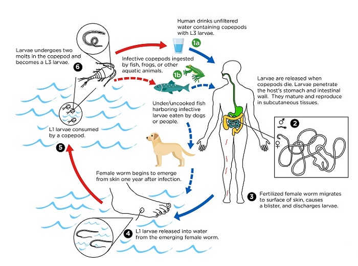
Dracunculus medinensis life cycle (Source: CDC)
Pathogenesis, Pathology of Dracunculus medinensis
The third-stage larvae of Dracunculus medinensis are not pathogenic. They do not produce any pathological lesions in humans.
The adult female parasite is pathogenic. It produces a fluid-filled blister at the site from where the female worm penetrates the host skin to release larvae – when it comes in contact with the skin. Dracunculus medinensis also produces diffusible toxins which may result in conditions such as vomiting, mild fever, urticaria, dyspnoea, and occasional fainting.
Host immunity against Dracunculus medinensis
There is no host immunity induced against Dracunculus medinensis infection, which is unusual. Individuals have been infected several times.
Clinical Manifestations of Dracunculus medinensis
The pre-patent period of Dracunculus medinensis infection is around 10 to 14 months. The parasitic infection is asymptomatic until the gravid female worm reaches the surface of the skin – ready to discharge the larvae.
Clinical manifestation of Dracunculus medinensis begins with a stinging papule developing into a blister. These blisters are usually found on the lower parts of the legs such as between metatarsal bones, soles of feet, and ankles. Blisters can also be found, less frequently, in the hand, trunk, calf, thigh, arm, buttock, knee joint, head, neck, and female breast.
An intense burning pain accompanies blister formation that can be relieved by immersion of the affected area in the water.
In some patients, Dracunculus medinensis infection may manifest as urticaria, diarrhea, dyspnoea, giddiness, nausea, vomiting, and marked burning sensations.
As the lesion vesiculates over the next few days, the blister ruptures producing a painful ulcer. The female worm is often visible in the ulcer’s opening while the larvae can be collected intermittently when the ulcer is douched with fluid. After the female parasite dies in the next few weeks, it is slowly absorbed into the tissue. The ulcer then eventually heals.
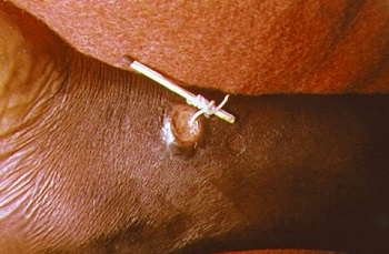
traditional extraction method of female Dracunculus medinensis (Source: Wikipedia)