Enterobius vermicularis - Introduction, Classification, History, Habitat, Morphology
Introduction to Enterobius vermicularis
Enterobius vermicularis is a common intestinal parasitic nematode (roundworm) that causes pinworm infection or enterobiasis. It is also known as threadworm, pinworm, or seatworm. This infection is most common in children and is transmitted via the fecal-oral route.
Although the parasite occurs exclusively in humans, infections have been reported from bonnet macaque.
Classification of Enterobius vermicularis
The classification of Enterobius vermicularis is as follows:
Kingdom: Animalia
Phylum: Nematoda
Class: Chromadorea
Order: Rhabditida
Family: Oxyuridae
Genus: Enterobius
Species: vermicularis
History of Enterobius vermicularis
The life cycle of the Enterobius vermicularis parasite was first described in 1865 by Leuckart.
Habitat of Enterobius vermicularis
The gravid female Enterobius vermicularis reside in the large intestine. The pinworm remains attached to the vermiform appendix, mucosa of the caecum, and adjacent parts of the large intestine.
Morphology of Enterobius vermicularis
The significant morphological forms of Enterobius vermicularis include:
Adult worm
Egg (infective form)
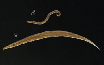
Male and female Enterobius vermicularis (Source: ResearchGate)
Adult worm
adult Enterobius vermicularis worms are white, small, spindle-shaped and thread-like
visible to the naked eye
absence of true buccal capsule
has a pair of wing-like expansions called cervical alae at the anterior end
presence of a conspicuous double-bulb esophagus
Male
adult Enterobius vermicularis male measures 2 to 5 mm long and 0.2 mm thick
has a sharply curved but blunt posterior end with a conspicuous terminal copulatory spicule
die immediately after fertilizing females, so rarely seen in samples
Female
adult Enterobius vermicularis female measures 8 to 13 mm long, and 0.5 mm thick
straight with a sharply pointed posterior end
presence of paired and T-shaped reproductive organs
uteri fill up the entire length of the female parasite
each gravid female’s uteri can contain 4,600 to 16,900 eggs with an average of 11,000
lives upto 2 months
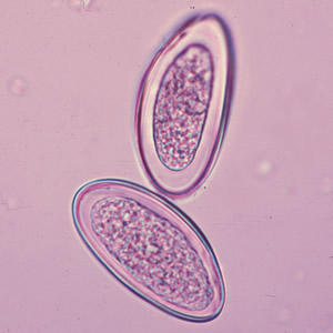
Eggs of Enterobius vermicularis (Source: ResearchGate)
Infective form
Eggs
eggs of Enterobius vermicularis are an infective form of the parasite
are translucent and non-bile stained
measures 50 μm to 60 μm in length and 20 μm to 32 μm in breadth
typically planoconvex – with one side convex and the other side flattened
the eggshell is transparent and hyaline
each egg encases a coiled larva or a developing embryo
small size and transparency of the eggs make them invisible to the naked eye except if eggs are clumped into thousands