Enterobius vermicularis - Laboratory Diagnosis, Treatment, Prevention, Control
Laboratory diagnosis of Enterobius vermicularis
Laboratory diagnosis of Enterobius vermicularis is done via demonstration of pinworm eggs in perianal or perineal scrapings and less commonly the adult worm.
Sample
The samples collected for the diagnosis of Enterobius vermicularis include:
anal swab
stool
urine (less common)
vaginal smear (less common)
Microscopy
Simple microscopy of less common samples such as stool, urine, or vaginal smears can also be done. Samples may also contain the adult worm itself which is also diagnostic of the pinworm infestation,
Microscopy of anal swabs is done to demonstrate the Enterobius vermicularis eggs by following the scotch-tape swab method.
 concentrated stool smear.jpg)
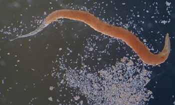
Male Enterobius vermicularis from a formalin-ethyl acetate (FEA) concentrated stool smear (Source: ResearchGate)
Scotch-tape swab method
The scotch tape swab method is a simple and effective method for the diagnosis of Enterobius vermicularis infection.
It is also called the transparent adhesive tape test or Cellophane-swab technique and is a quick, cost-effective, and painless test. Multiple samples (for three consecutive days) may be collected at home early in the morning before and submitted later to the laboratory/doctor's office.
In this method, a sample from the skin around the anus is collected and examined. The sample must be retrieved before the patient has gotten out of bed, used the bathroom, or taken a bath.
the sticky side of a piece of clear tape (such as Scotch tape) is applied briefly to the skin around the patient’s anus
any eggs present on the skin will stick to the tape
the sticky side of a piece of the tape is pressed to a microscope slide and viewed under a microscope
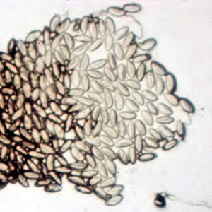
Eggs of Enterobius vermicularis in a cellulose-tape preparation (Source: ResearchGate)
Stool microscopy
Among numerous stool microscopy methods, direct wet mount microscopy is done for the diagnosis of Enterobius vermicularis.
Direct wet mount microscopy
the wet mount for Enterobius vermicularis is prepared by mixing stool sample with saline, iodine, or lactophenol cotton blue (LPCB)
enterobiasis is confirmed by the demonstration of the pinworm eggs
Stool concentration
the formalin-ether method or salt flotation of stool is used for the concentration of the specimen for the detection of eggs of Enterobius vermicularis
Adult Enterobius vermicularis surrounded by eggs (Source: ResearchGate)
Treatment of Enterobius vermicularis
The treatment of Enterobius vermicularis is done by administration of drugs such as Pyrantel pamoate, mebendazole, albendazole, piperazine, and pyrvinium pamoate.
Prevention, and Control of Enterobius vermicularis
Prevention, and control of Enterobius vermicularis are done by:
Individual prophylaxis
Individual prophylaxis of Enterobius vermicularis is done by
improved personal hygiene such as proper washing of hands with soap after defecation and before eating food
treatment of water before drinking such as boiling, filtering the water used in a 0.22-micrometer membrane, iodination with tetracycline hydro per iodide
drinking bottled water while traveling to areas endemic to enterobiasis
if salad is to be consumed, treat the vegetables with acetic acid or vinegar for 15 minutes
not performing sexual acts that involve fecal-oral contact
Community prophylaxis
Community prophylaxis of Enterobius vermicularis is done by
improvement of the water management system to avoid fecal contamination
improvement of sanitation by installing latrines for proper disposal of human feces