Leishmania tropica complex - Laboratory diagnosis, Treatment, Prevention, Control
Laboratory diagnosis of Leishmania tropica complex
The laboratory diagnosis of the Leishmania tropica complex begins with the collection of samples.
Specimen
The specimen for lab diagnosis of Leishmania tropica complex includes
the outer edge of the ulcer aspirates
a swab of ulcer margin
skin biopsy from the ulcer margin
Microscopy
stains such as Leishman stain, Giemsa stain, Brown-Hopps stain, or Wright stain can be used for microscopy
presence of amastigotes confrims the diagnosis
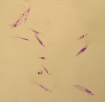
Image: Leishmania tropica Complex microscopy (Source: Wikipedia)
Culture
This method of culture is followed in cases of Leishmaniasis recidivans and in cases suspected of cutaneous leishmaniasis but the parasites are not seen in direct microscopy of Leishmania tropica complex.
In-vitro culture
media used for in-vitro cultures includes NNN, Schneider Drosophila medium, or any biphasic medium
samples are inoculated into the water of condensation or into the fluid of a liquid medium
incubated at 22° C - 26° C for 1-4 weeks
at the end of each week, a drop of culture is microscopically examined for promastigotes
motile promastigotes can be observed in positive cultures
has a sensitivity of 75%
Animal inoculation
laboratory animals such as Chinese and golden hamsters are inoculated intraperitoneally for animal inoculation
skin biopsy, lesion aspirations are stained and visualized under a microscope
Serodiagnosis
Immunodiagnosis/serodingaosis methods for Leishmania tropica complex diagnosis such as Complement Fixation Test (CFT), Indirect Immuno-fluorescent (IFA), Enzyme-Linked Immunosorbent Assay (ELISA), and Direct Agglutination test (DAT) are not used in the diagnosis of cutaneous leishmaniasis due to low level of leishmanial antibodies in the serum.
Aldehyde test of Napier
Although a very useful test for the diagnosis of Leishmania donovani, Aldehyde test of Napier is negative and not useful in cutaneous leishmaniasis.
Leishmania skin test (Montenegro test)
Leishmania tropica complex infection can be diagnosed by the Leishmania skin test (Montenegro test).
delayed hypersensitivity test and is helpful
the antigen used in this method is obtained from killed cultured promastigotes of Leishmania tropica
indicates positive results just a few days after the infection
positive skin test results are also seen in treated cases as well as Leishmaniasis recidivans
negative in cases of Diffuse cutaneous leishmaniasis
0.2 ml of Leishmania antigen is injected intradermally and read after 48-74 hours
induration and erythema of 5mm diameter or larger are seen in positive cases
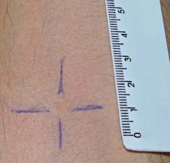
Image: Leishmania skin test (Source: ResearchGate)
Treatment of Leishmania tropica complex
The treatment for Leishmania tropica complex infection involves:
in endemic areas, treatment is not started until the ulcers develop as it induces the development of host immunity
if the ulcers have been persisting for more than 6 months or the ulcers are disfiguring, they are treated with antimonial drugs
Drugs used include:
Pentavalent antimonials
megalumine antimonate
sodium stibugluconate solution
Pentamidine
pentamidine isethionate
pentamidine dimethane
Amphotericin B
Miltefosine
Interferon
Prevention, Control of Leishmania tropica complex
The prevention, control of Leishmania tropica complex can be done by following methods:
reduction of sand-fly population by using insecticides to reduce the source of Leishmania tropica complex
evade contact with potential animal vectors such as dogs, foxes, and rodents
use of bed nets, window nets, or insect repellents