Microsporidia - Clinical Manifestation, Prognosis, Epidemiology, Reservoir, Lab diagnosis, Treatment
Clinical manifestation of Microsporidia
Microsporidia is an opportunistic parasite that usually affects immunocompromised individuals while infection in immunocompromised individuals is uncommon. The clinical manifestation of Microsporidia includes
intestinal microsporidiasis
disseminated microsporidiosis
ocular infection
musculoskeletal infection
Intestinal microsporidiasis
Enterocytozoon bieneusi causes 90% of intestinal microsporidiosis
Encephalitozoon intestinalis is responsible for 10% of cases
intestinal microsporidiasis is usually seen in patients with AIDS (CD4 <100mm3)
characteristics include protracted debilitating chronic diarrhea (lasts several months), malabsorption, and wasting
the mortality rate is high as 56% in individuals with diarrhea
Disseminated microsporidiosis
Encephalitozoon intestinalis causes the dissemination of microsporidiosis
dissemination can involve ocular, genitourinary, and respiratory tracts
in advanced cases, ocular infections, respiratory infections, and renal failure occurs
Enterocytozoon bieneusi causes acalculous cholecystitis
Encephalitozoon hellem and Encephalitozoon cuniculi can produce clinical manifestations such as keratoconjunctivitis, respiratory tract infection, genitourinary tract infection as well as disseminated infections
Ocular infection
Vittaforma corneae, Nosema connori, and Nosema ocularum cause ocular infection
this condition manifests as a foreign body sensation, eye redness, excessive lacrimation, and blurred or decreased vision
cornea infection is caused by Microsporidium ceylonensis and Microsporidium africanum
Musculoskeletal infection
clinical syndromes include myalgia, fever, generalized muscle weakness
musculoskeletal infection is caused by Pleistophora and Trachipleistophora hominis
Image: Transmission electron micrograph of a microsporidian spore with an extruded polar tubule inserted into a eukaryotic cell (Source: CDC)
Prognosis of Microsporidia
In severely immunocompromised individuals and in cases of disseminated microsporidiosis, the prognosis is poor.
Epidemiology of Microsporidia
Epidemiologically, Microsporidia is cosmopolitan in distribution. Human infections have been reported in both developed and underdeveloped nations- in immunocompromised as well as immunocompetent individuals.
Reservoir, Source, Transmission of Microsporidia
Spores are the infectious stage of Microsporidia.
Transmission occurs via inhalation or ingestion of spores.
Laboratory Diagnosis of Microsporidia
The laboratory diagnosis of Microsporidia is based on the demonstration of spores in the specimen.
Sample
stool
urine
mucous smears
biopsy/autopsy tissue
Microscopy
stains including modified trichrome stains (chromotrope 2R), fluorochrome stains (calcofluor white, Uvitex 2B) can be used for microscopy
if a modified trichrome stain is used the parasite appears small, oval, refractile spores with bright pinkish-red walls
a belt-like stripe, stained pink, may also be visible in the middle of the spore
the spore retains dark violet and the belt-like stripe is enhanced by the rapid Gram chronotrope method
other stains such as Warthin-Starry silver stain, Brown Brenn Gram stain, Giemsa stain, and trichrome clue can be used to detect spores microscopically in biopsy or autopsy tissue samples
use of monoclonal and/or polyclonal antibodies in a direct fluorescent method can demonstrate spores as well as extruded polar tubules
Transmission electron microscopy is the gold standard for the diagnosis and identification of Microsporidia but is expensive and time-consuming
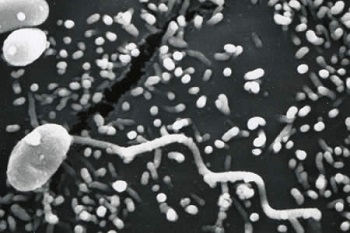
Image: Scanning electron micrograph of a microsporidian spore with an extruded polar tubule inserted into a eukaryotic cell (Source: CDC)
Serodiagnosis
There are no serological tests for laboratory diagnosis of Microsporidia.
Molecular diagnosis
PCR can be used for the molecular diagnosis and species identification of Microsporidia.
Treatment of Microsporidia
Albendazole can be administered for the treatment of intestinal, disseminated, and ocular microsporidiosis