Microsporidia - Introduction, Classification, Morphology, Life Cycle, Pathogenesis, Pathology
Introduction of Microsporidia
Microsporidia are spore-forming, obligate eukaryotic intracellular parasites. The spores contain extrusion apparatus which has a coiled polar tube ending in an anchoring disc at the apical end. They have a unique mechanism to infect host cells such as:
resistant spores which vary in size depending on the species
has a polar tubule or polar filament while a unique organelle is present on the side of the spore
The phylum Microspora contains more than 1,200 species from 143 genera. At least 13 species have been known to cause disease in humans:
Enterocytozoon bieneusi
Encephalitozoon hellem
Encephalitozoon intestinalis
Encephalitozoon hominis
Pleistophora
Trachipleistophora hominis
Trachipleistophora anthropophthera
Nosema connori
Nosema ocularum
Brachiola vesicularum
Vittaforma corneae
Microsporidium ceylonensis
Microsporidium africanum
Classification of Microsporidia
The classification of Microsporidia can be done by:
(unranked): Obazoa
(unranked): Opisthokonta
Clade: Holomycota
Kingdom: Fungi
Subkingdom: Rozellomyceta
Phylum: Rozellomycota
Class: Microsporidea
History of Microsporidia
Historically, they were classified as protozoans or protists and now it has been to be fungi or a sister group of fungi.
Habitat of Microsporidia
Microsporidia inhabit the small intestine of human hosts.
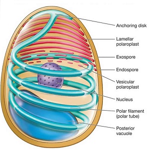
Figure: Microsporidia morphology (Source: Health Jade)
Morphology of Microsporidia
Spore is one of the important morphological features of Microsporidia.
Spores
spore is the infective form of the parasite
size of the spores depends on the species of Microsporidia
Encephalitozoon spp, Vittaforma corneae, Nosema spp – 1.5 μm to 4 μm
Encephalitozoon biemeusi – 0.8 μm to 1.4 μm
resistant forms of Microsporidia
can survive several months in the environment
Life Cycle of Microsporidia
Humans acquire microsporidiosis after ingestion or inhalation of Microsporidia spores, thus starting its life cycle.
The polar tubules erupt from the apical end of the spore which injects the infective sporoplasm into the host cell
the sporoplasm multiplies inside the cell either by merogony (binary fission) or schizogony (multiple fission)
merogony and schizogony can take place directly inside the host cell cytoplasm (in Encephalitozoon bieneusi) or inside a parasitophorous vacuole (in Encephalitozoon intestinalis)
Microsporidia undergo sporogony to develop into mature spores
maturation of spores also includes thickening of cyst wall
the host cells after completely filled with spores, stretch and finally ruptures to release the spores
thick spore wall is able to protect the parasite during unfavorable environmental conditions
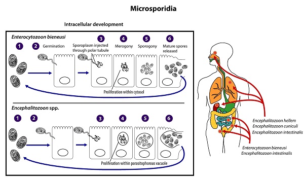
Figure: Microsporidia life-cycle (Source: CDC)
Pathogenesis, Pathology of Microsporidia
Microsporidia are invasive intracellular parasites with Encephalitozoon intestinalis being more invasive.
they invade the intestinal mucosa and are usually found only in enterocytes
in the intestinal mucosa, infected enterocytes are distributed in patches
the infected enterocytes show pathological changes such as villous atrophy, elongated crypts, and depletion of goblet cells
in AIDS patients, the biliary tract is also infected which results in cholangitis and cholecystitis
cells of lamina propria, macrophages, and enterocytes are also infected
disseminated infections can also occur by invading the small intestine, large intestine, gallbladder, urinary tract, respiratory tract, and epithelial cells of the respiratory and urinary tract