Naegleria fowleri - Life Cycle, Pathogeneis, Pathology, Host Immunity, Treatment, Control
Life Cycle of Naegleria fowleri
The life cycle of Naegleria fowleri completes in man through a cycle of asexual generation.
infection is acquired by swimming or diving in warm water contaminated with the amoebae which are neurotrophic
the point of entry in the host is the nose from where it invades the olfactory mucosa and olfactory bulbs
from there they penetrate the submucosal nervous plexus, invade the cribriform plate and eventually reach the subarachnoid space
the host CSF contains glucose and protein which supports the growth and multiplication of Naegleria fowleri
Also, the high concentration of oxygen present in the CSF and brain of the host promotes the growth as well as multiplication of the amoebae
through the foramen of Luschka and Magendie, the trophozoites enter the ventricular system and reach the choroid plexus where they destroy the ependymal layer of the third, fourth, and lateral ventricles resulting in acute ependymitis
by the process of promitosis, the trophozoites multiply
during promitosis, an intact nuclear membrane is present which can be demonstrated by an electron microscope
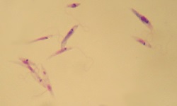
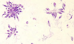
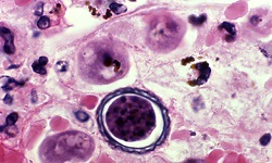
Naegleria fowleri trophozoites are found in pathogenic CNS lesions which are located mainly around blood vessels (Colour plate-III J) while cysts are not seen as the rapid infection results in quick death- before trophozoites are able to encyst
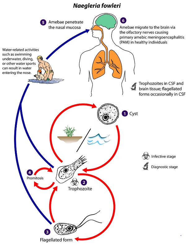
Fig: life cycle of N. fowleri (Source: CDC)
Pathogeneis, Pathology of Naegleria fowleri
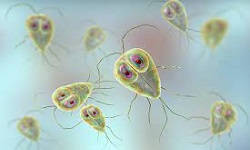
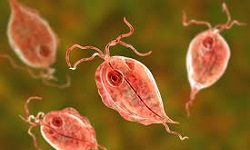
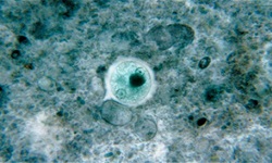
Naegleria fowleri trophozoites are neurotrophic, and phagocytic and are able to ingest RBCs as well as brain tissues
in the host brain tissue, the amoebae produce a food cup called amebostome into which cytopathic enzymes such as phospholipases, hydrolases, and lysosomal enzymes are secreted
* These enzymes from Naegleria fowleri are responsible for the destruction of neural tissues in the brain.
In addition, resistance to the complement system of the host and the ability to migrate is the virulence factor of the amoebae
they are able to invade the CNS, and through the olfactory neuro-epithelium, fascicles of the olfactory nerve, reach the subarachnoid space and brain
they subsequently disseminate to the other areas of CNS from the subarachnoid space
Naegleria fowleri produces acute hemorrhagic necrotizing meningoencephalitis associated with moderate purulent exudates. This is found mainly in the cerebellum, brain stem, and base of the brain while the olfactory mucosa and olfactory bulbs are the most commonly affected areas.
As the brain suffers severe edema, the CSF pressure is elevated resulting in cerebellar or uncal herniation
Host Immunity of Naegleria fowleri
As the course of Naegleria fowleri infection is rapid and fulminant, patients suffering from primary amoebic meningoencephalitis (PAM) die usually die within 5-10 days. Thus, the specific antibody level at which they can be detected is not produced in the serum and the host immunity is relatively absent.
Treatment of Naegleria fowleri
Only a few cases have survived infection from Naegleria fowleri and all these cases were diagnosed and treated in the early stages. Drugs of choice include:
Amphotericin B
Miconazole
Sulfisoxazole
Phenothiazine
Rifampin
Prevention, Control of Naegleria fowleri
The prevention, control of Naegleria fowleri are done as follows:
avoid contact with stagnant water, especially where Naegleria fowleri has been detected in its sources
hyper chlorination of swimming pool water does not kill the amoebae