Plasmodium ovale - Introduction, Classification, History, Habitat, Morphology
Introduction of Plasmodium ovale
The genus Plasmodium causes the vector-borne disease malaria. These parasites show an alternation of generation accompanied by an alternation of hosts. In the human host, the asexual cycle (schizogony) takes place inside the erythrocytes while the sexual cycle (sporogony) takes place in the mosquito host.
Typically, the infected erythrocytes produce pigments that are visualized by light microscopy.
Plasmodium vivax, P. ovale, and P. malariae belong to the subgenera Plasmodium while P. falciparum belongs to the subgenus Laverania.
Plasmodium ovale is the causative agent of ovale or mild tertian malaria. They also cause relapse like P. vivax.
Classification of Plasmodium ovale
Kingdom: Chromista
Subkingdom: Harosa
Infrakingdom: Halvaria
Superphylum: Alveolata
Phylum: Apicomplexa
Class: Aconoidasida
Order: Haemospororida
Family: Plasmodiidae
Genus: Plasmodium
Species: P. ovale
History of Plasmodium ovale
MacFie and Ingram, in 1917, first described Plasmodium ovale in Ghana. In 1918, Stephens discovered the malaria parasite in a soldier from East Africa and termed the name Plasmodium ovale.
Habitat of Plasmodium ovale
Plasmodium ovale typically infects young Red Blood Cells.
Morphology of Plasmodium ovale
The various morphological forms of Plasmodium ovale depend on the host the parasite is residing in i.e. the diagnostic form or the infective form.
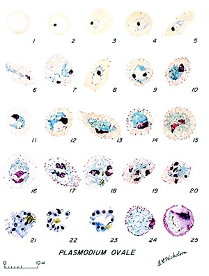
Figure: Development of the erythrocytic stages of Plasmodium ovale. 1, normal red cell; 2 to 5, young trophozoites; 6 to 11, growing trophozoites; 12 and 13, nearly mature and mature trophozoites, respectively; 14 to 20, developing schizonts; 21 and 22, mature schizonts; 23, developing gametocyte; 24, mature macrogametocyte; 25, mature microgametocyte (Source: journals.asm.org)
Diagnostic forms in humans
The diagnostic forms of Plasmodium ovale found in the human host include
Early trophozoite (ring form)
Late trophozoite (trophozoite form)
Schizont
Gametocytes
Early trophozoite (ring form)
are relatively large and occur in peripheral blood
have a delicate blue-stained ring of cytoplasm with a red chromatin dot
in some cases, two red chromatin dots can be found separated or closed together
sometimes, two early trophozoites can be found in a single infected RBC
the cytoplasm of Plasmodium ovale stains deep blue than P. vivax
also, the cytoplasm of Plasmodium ovale is smaller, thicker, and heavier than that of P. vivax
Late trophozoite (trophozoite form)
smaller and compact
consists of coarse pigment granules and an inconspicuous vacuole
lacks amoeboid form
Schizont
mature schizonts almost fill their host cells (three-fourths)
young schizonts are small and compact with few chromatin masses and yellow-coarse coarse pigments
mature schizont contains around 10 merozoites which are arranged in the form of an irregular rosette
Plasmodium ovale merozoites are larger than P. malariae
Gametocytes
There are two types of gametocytes- macrogametocytes, and microgametocytes. These gametocytes are smaller and not abundant as P. vivax. They take a longer time to appear in peripheral blood than other malaria parasites.
Macrogametocytes
they are round or oval, compact, and filled with enlarged erythrocytes
smaller nucleus with a compact mass of chromatin
the fine granules are arranged in small masses and occur near the periphery
the cytoplasm stains blue while chromatin pigments deep red and violet
Microgametocytes
smaller than macrogametocytes
oval or round with a large nucleus
chromatin granules are arranged to form a spindle
cytoplasm, which is dark blue, contains dark, coarse hemozoin pigments distributed throughout the cytoplasm
do not occupy the entire host RBC
* Infected host erythrocytes
the infected host is enlarged, oval, fimbriated
develops Schuffner’s dots or James’ dots, which is a characteristic stippling and can be demonstrated with Romanowky’s stain of blood smear
electron microscopy reveals surface invaginations surrounded by small vesicles
Infective form
The infective form of Plasmodium ovale for humans is the sporozoites.
Sporozoites
measures 11μm to 12μm
elongated with pointed ends