Plasmodium vivax -Introduction, History, Habitat, Morphology, Culture
Introduction of Plasmodium vivax
The genus Plasmodium causes the vector-borne disease malaria. These parasites show an alternation of generation accompanied by an alternation of hosts. In the human host, the asexual cycle (schizogony) takes place inside the erythrocytes while the sexual cycle (sporogony) takes place in the mosquito host.
Typically, the infected erythrocytes produce pigments that are visualized by light microscopy.
Plasmodium vivax, P. ovale, and P. malariae belong to the subgenera Plasmodium while P. falciparum belongs to the subgenus Laverania.
Plasmodium vivax is the causative agent of benign tertian or vivax malaria. The characteristic of this type of malaria is the occurrence of true relapses.
History of Plasmodium vivax
The first description of Plasmodium vivax came in 1880 by Laveran but he failed to distinguish the species. Six years later, in 1886, Golgi described the parasite as a distinct species of Plasmodium. It was then given the name Haemamoeba vivax in 1890 by Grassi and Feletii.
Habitat of Plasmodium vivax
Plasmodium vivax at various stages is found inside the parenchymal cells of the liver and the red blood cells (erythrocytes). During the erythrocytic schizogony cycle, all the forms of the malaria parasite are found in the peripheral blood.
Morphology of Plasmodium vivax
The various morphological forms of Plasmodium vivax are dependent on the host the parasite is residing in i.e. the diagnostic form or the infective form.
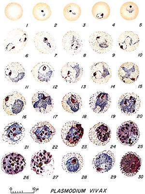
Figure: P. vivax morphological forms- 1: Normal red cell; 2-6: Young trophozoites (ring stage parasites); 7-18: Trophozoites; 19-27: Schizonts; 28 and 29: Macrogametocytes (female); Microgametocyte (male) (Source: U.S. Department of Health, Education and Welfare; 1971)
Diagnostic form
The diagnostic forms of Plasmodium vivax found in the human host include
Early trophozoite (ring form)
Late trophozoite (trophozoite form)
Schizont
Gametocytes
Early trophozoite (ring form)
are relatively large and occur in peripheral blood
have a delicate blue-stained ring of cytoplasm with a red chromatin dot
in some cases, two red chromatin dots can be found separated or closed together
sometimes, two early trophozoites can be found in a single infected RBC
Late trophozoite (trophozoite form)
are large and amoeboid
consists of abundant chromatin, dark pigment granules, and a vacuole which is characteristic
during this form, a mass of brown hemozoin almost fills the RBC
Schizont
they are large, round, and irregular
occupy the entire infected which has also been enlarged
hemozoin which almost fills the erythrocytes are present in one or two clumps
mature schizont contains around 12 to 24 merozoites
Gametocytes
There are two types of gametocytes in Plasmodium vivax - macrogametocytes, and microgametocytes.
Macrogametocytes
measures 10μm in diameter
they are round or oval, compact, and filled with enlarged erythrocytes
smaller nucleus with a compact mass of chromatin
the fine granules are arranged in small masses and occur near the periphery of Plasmodium vivax
the cytoplasm stains blue while chromatin pigments deep red and violet
Microgametocytes
smaller than macrogametocytes
oval or round with a large nucleus
chromatin granules are arranged to form a spindle
cytoplasm, which is dark blue, contains dark, coarse hemozoin pigments distributed throughout the cytoplasm
do not occupy the entire host RBC
* Infected host erythrocytes
the infected host erythrocytes are enlarged, irregular in shape, and pale in color
develops Schuffner’s dots, which is a characteristic stippling and can be demonstrated with Romanowky’s stain of blood smear
electron microscopy reveals surface invaginations surrounded by small vesicles
Infective form
The infective form of Plasmodium vivax for humans is the sporozoites.
Sporozoites
measures 10μm to 14μm
sickle-shaped
presence of a single nucleus
found in the salivary gland of the mosquito
Culture of Plasmodium vivax
In-vitro
The culture of Plasmodium vivax for routine diagnosis is not done due to limited success as it is still in the experimental stage.
However, it has been cultured in in-vitro in human erythrocytes and is done in media with of following properties:
consists of RPMI 1940 medium
an overlay of medium with human serum
a thin layer of stationary human blood cells
maintained with 7% carbon dioxide in the atmosphere
maintained with 1% to 5% oxygen in the atmosphere
Laboratory animals
The Plasmodium vivax malaria parasite has been cultured in several species of primates such as monkeys.