Taenia saginata - Introduction, Classification, History, Habitat, Morphology
Introduction of Taenia saginata
Taenia saginata is a large zoonotic tapeworm causing intestinal taeniasis in humans. It is also known as the beef tapeworm. Infection is acquired orally by ingesting raw or undercooked beef containing the larvae (Cysticercus bovis) of the parasite.
Cattles are intermediate hosts where larval development takes place while the definitive host of Taenia saginata is the human host harboring the adult tapeworm. The parasite does not cause cysticercosis and the infection is harmless and asymptomatic in most cases.
Taenia saginata strongly resembles other human tapeworms such as Taenia asiatica and Taenia solium, in structure and biology. However, Taenia saginata can be differentiated on the basis of the following properties:
typically larger and longer than Taenia asiatica and Taenia solium
more proglottids, more testes, and higher branching of the uteri
lacks an armed scolex
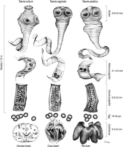
Fig: Morphological differences between Taenia spp (Source: ResearchGate)
Classification of Taenia saginata
Kingdom: Animalia
Phylum: Platyhelminthes
Class: Cestoda
Order: Cyclophyllidea
Family: Taeniidae
Genus: Taenia
Species: T. saginata
History of Taenia saginata
In 1782, Goeze differentiated Taenia saginata from T. solium. Leuckart, in 1863, demonstrated cattle to be the intermediate host of the tapeworm.
Habitat of Taenia saginata
In the definitive host, the adult tapeworm can be found in the small intestine (upper jejunum). The parasite remains attached to the intestinal mucosa with the help of suckers.
Morphology of Taenia saginata
Adult worm
white, ribbon-like, flattened, and segmented worm
measures 4 meters to 10 meters but sometimes may reach upto 15 meters
contains head (scolex), neck, and body (strobila) which consists of a chain of segments (proglottids)
Scolex
quadrate in shape
measures 2mm in diameter
contains four cup-shaped muscular suckers
suckers may be pigmented
hooks or rostellum is absent
Neck
short and fragile
measures 3mm to 7mm in length
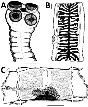
Fig: T. saginata - A - scolex, B - Gravid proglottid, C - mature proglottid, (Source: ResearchGate)
Strobila
measures 4 meters to 10 meters but sometimes may reach upto 15 meters
consists of 1000-2000 proglottids or segments
the proglottids of Taenia saginata are arranged in a linear sequence of immature, sexually mature, and gravid
each proglottid consists of numerous testes and bi-lobed ovary
on the lateral part of each segment, common genital pores are present with alternative irregularly between the right and left margin
sexually mature segments are opaque, and white and measure 2cm in length and 5mm to 7mm n breadth
gravid segments are rectangular measuring 20mm by 5mm to 7mm and are found in the posterior part of the parasite
as gravid segments uterus is fully developed, it consists of 15-30 lateral branches
These gravid proglottids routinely break off from the strobila and are passed along with the feces.
The proglottids of Taenia saginata are different from that of T. solium as Taenia saginata:
has twice the number of testes i.e. 300-400 testes than present in T. solium
have developed vaginal sphincter
lacks accessory ovarian lobe
typically larger and longer than Taenia asiatica and Taenia solium
more proglottids, more testes, and higher branching of the uteri
lacks an armed scolex
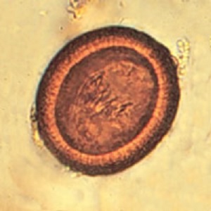
Image: Taenia egg (Source: cych.org)
Egg
round or oval in shape
measures 33μm to 43μm in diameter
bile stained
each egg has two membranes or shells- an outer membrane and an inner membrane
* outer shell, also called the egg capsule, is thin and transparent and is present when passed fresh from proglottids but is absent in eggs from old feces
* inner shell, also called embryophore, is a thick, brown roughly structured wall enclosing the embryo
* embryo (oncosphere) measures 14μm to 20μm in diameter, contains six hooklets (hexacanth embryo), and is only infective to cattle
since eggs are similar in size and shape to that of T. solium, Echinococcus, and Multiceps, light microscopy cannot identify Taenia saginata eggs
Infective Form
Cysticercus bovis
the cysticerci larvae (Cysticercus bovis) is the infective form for humans
oval in shape measuring 5mm x 10mm
has a translucent cyst filled with a clear fluid
contains opaque invaginated protoscolex (future scolex)
they can be found in any organ but is much more common in the heart and muscles of the cattle
this stage occurs only in cattle and not the humans