Toxoplasma gondii - Introduction, Classification, History, Habitat, Morphology, Culture
Introduction of Toxoplasma gondii
Toxoplasma gondii is an obligate intracellular parasite causing toxoplasmosis in humans. They are the only protozoans that are infectious to man in all the stages- tachyzoite, tissue cyst, and oocyst.
The definitive host is the feline while the bird or other mammals are the intermediate hosts. Merogony occurs in both definitive hosts and intermediate hosts. The sporogony and schizogony take place in the intestine of the definitive host.
Classification of Toxoplasma gondii
The classification of Toxoplasma gondii is done as:
Kingdom: Chromista
Superphylum: Alveolata
Phylum: Apicomplexa
Class: Conoidasida
Order: Eucoccidiorida
Family: Sarcocystidae
Subfamily: Toxoplasmatinae
Genus: Toxoplasma
Species: T. gondii
History of Toxoplasma gondii
Historically, Toxoplasma gondii was first discovered in 1908 by Nicolle and Manceaux in the spleen, liver, and blood of the North American rodent Ctenodactylus gondii. It was named Toxoplasma gondii in 1909. The coccidia was found to be infecting a man from Darjeeling. It was later found to be infecting a child congenitally in 1937 by Paige, Wolf, and Cowan.
Only in 1970 was the Toxoplasma gondii life cycle fully described with confirmation that cats are definitive hosts are other warm-blooded animals such as birds and mammals are intermediate hosts.
Habitat of Toxoplasma gondii
Since Toxoplasma gondii is an obligate intracellular parasite, they habitat by infecting reticuloendothelial cells and other nucleated host cells such as muscle and intestinal epithelium.
Morphology of Toxoplasma gondii
The morphological forms of Toxoplasma gondii include:
Diagnostic forms
The diagnostic forms of Toxoplasma gondii include tachyzoites and tissue cysts which are important diagnostic forms.
Tachyzoites
observed during the acute stage of infection
tachyzoites are active, multiplying trophozoites
oval to crescent shape with a rounded posterior end and pointed anterior end
the ovoid nucleus lies in the posterior end of tachyzoites
capable of invading all types of mammalian cells (except erythrocytes)
can be found in any organ but most commonly in skeletal muscle, cardiac muscle, and brain
once inside the host cell, they undergo endodyogeny which is multiplication within a vacuole
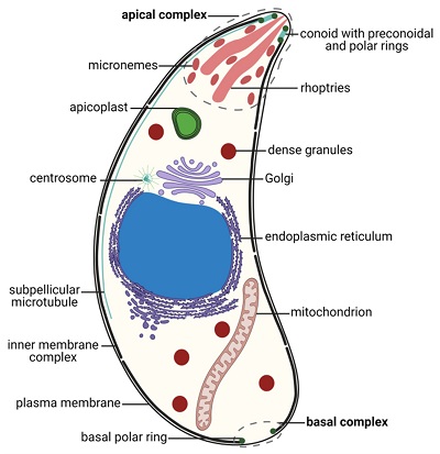
Figure: Toxoplasma gondii tachyzoite morphology (Source: MDPI)
Tissue Cyst, Bradyzoites
resting form of the parasite
tissue cyst, bradyzoites occur during the chronic stage of Toxoplasma gondii infection
spherical with size varying from 40mm to 50mm
each cyst is surrounded by an eosinophilic, weakly PAS-positive cyst wall
inside each cyst, hundreds of trophozoites called bradyzoites which are strongly PAS-positive are present
the bradyzoites, which are smaller than tachyzoites, undergo multiplication by endodyogeny
cysts extremely resistant to host defenses
tissue cysts can be found in any organ of the host but are most common in brain, skeletal, and heart muscles
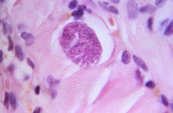
Image: hematoxylin and eosin-stained (H&E) photomicrograph of a Toxoplasma gondii tissue cyst with bradyzoites in human muscle tissue (Source: lecturio)
Oocyst
oocyst stage occurs in cats and other felines
oval measures 10 μm to 12 μm in diameter
each oocyst is enclosed in a thick, resistant wall
the unsporulated oocysts which are excreted in the cat feces are non-infectious
after undergoing sporulation in the environment, the sporulated oocysts containing two sporocysts each containing four sporozoites, are infectious to humans
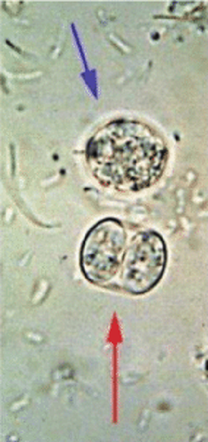
Image: Toxoplasma gondii oocyst - unsporulated (blue arrow), sporulating (red arrow) (Source: Reseachgate)
Culture of Toxoplasma gondii
Toxoplasma gondii can be cultured in-vitro as well as the laboratory animals.
In-vitro culture
Toxoplasma gondii can be cultured in vitro in normal murine alveolar and peripheral cell lines in tissue culture
they are yet to be successful to be cultured in a cell-free medium
Laboratory animals
laboratory animals such as mice and hamsters are intraperitoneally inoculated with Toxoplasma gondii