Cryptosporidium parvum - Introduction, Classification, History, Habitat, Morphology, Culture
Introduction of Cryptosporidium parvum
Cryptosporidium spp is a coccidian parasite which causes infection of the small intestine resulting in diarrhoea. Numerous species of Cryptosporidium are known to affect amphibians, fish, birds, and mammals while Cryptosporidium parvum is infectious to human beings.
Classification of Cryptosporidium parvum
Phenotypically, Cryptosporidium parvum can be classified on
Kingdom: Chromista
Superphylum: Alveolata
Phylum: Apicomplexa
Class: Conoidasida
Order: Eucoccidiorida
Suborder: Eimeriorina
Family: Cryptosporidiidae
Genus: Cryptosporidium
Species: parvum
History of Cryptosporidium parvum
Tyzzer, in 1907, was the first to describe the parasite obtained from the peptic glands of laboratory mice and named it Cryptosporidium. These coccidians were first thought to be non-pathogenic as only 15 reports of Cryptosporidium infections were reported in animals before 1975. However, in 1976, Cryptosporidium infection was reported in a three-year-old healthy girl in the USA.
Since then the Cryptosporidium infection has been diagnosed in individuals with AIDS or people undergoing immunosuppressive therapy.
Habitat of Cryptosporidium parvum
Cryptosporidium parvum inhabits the small intestine’s surface epithelial cells of villi or crypts of the small intestine.
The parasite may also inhabit the stomach, appendix, colon, rectum, and pulmonary tree.
Morphology of Cryptosporidium parvum
Cryptosporidium parvum exists in six different morphological forms:
Oocyst
sporozoite
trophozoite
meront
microgamont
macrogamont
Oocyst
diagnostic form excreted in human faeces
colourless, spherical to oval
Cryptosporidium parvum oocyst measures 4.5 μm to 6 μm in diameter
the cyst is surrounded by a 50 nm thin cyst wall which in turn consists of an electroluminescent middle zone surrounded by two electron-dense layers
does not stain with iodine and is acid-fast
each oocyst consists of up to four sporozoites
the sporozoites are slender, bow-shaped, and fusiform which remain parallel to each other
these sporozoites are released after partial digestion of oocyst
micropile polar granules are absent in Cryptosporidium parvum
oocyst, which sporulates inside the host, are excreted in small numbers in faeces
the severity of the infection and the number of oocysts excreted has no co-relation
oocysts are either thick-walled or thin-walled
thick-walled Cryptosporidium parvum oocysts are infectious to susceptible humans while thin-walled oocysts cause autoinfection in the same host
Sporozoite
Cryptosporidium parvum sporozoite is slender, crescent-shaped
measures 1.5 μm to 1.75 μm in diameter
pointed anterior end
the posterior end contains a rounded prominent nucleus
Trophozoite
Cryptosporidium parvum trophozoite is an intracellular transitional form of the parasite
round or oval
2μm to 2.5μm in diameter
each trophozoite consists of a large nucleus with/without a conspicuous nucleolus
Meronts
Cryptosporidium parvum meronts are crescent-shaped
measures 1μm to 5μm in diameter
rounded anterior and posterior ends
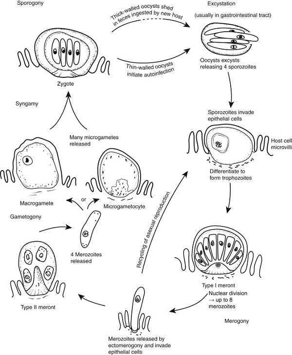
Figure: Cryptosporidium parvum developmental stages (Source: Science Direct)
Culture of Cryptosporidium parvum
Cryptosporidium parvum can be cultured in the media as well as the laboratory animals.
In media
Cryptosporidium parvum can be cultured in-vitro in tissue culture using a variety of cell lines such as monolayered human foetal lung, primary chick kidney, porcine kidney etc as well as in chick embryos
in cell lines, development takes place through type I meronts, type II meronts, sexual forms (microgamont, macrogamont), and oocysts
Laboratory animals
Cryptosporidium parvum is infectious to sucking mice and can be cultured in them.