Cryptosporidium parvum - Laboratory Diagnosis, Treatment, Prevention, Control
Laboratory diagnosis of Cryptosporidium parvum
Since cryptosporidiosis clinically resembles giardiasis, isosporiasis, and cyclosporiasis, the clinical diagnosis is difficult. However, differentiation can be made by the absence of blood, pus cells, and Charcot-Leyden crystals may possibly point toward cryptosporidiosis.
Identification and demonstration of Cryptosporidium parvum oocysts in the specimen are done for diagnosis of infection.
Sample
The samples collected for laboratory diagnosis of Cryptosporidium parvum include
stool
sputum
bronchial washings
duodenal or jejunal aspirations
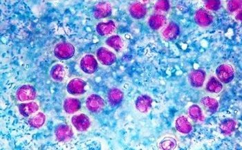
Figure: Cryptosporidium parvum microscopy after Kinuoun's stain (Source: YouTube)
Microscopy
Microscopic examination of the specimen after wet mount preparation and stained smear preparation is done to determine the presence/absence of oocysts.
Since Cryptosporidium parvum oocysts are excreted variably in the stool, multiple stool samples should be examined. Also, the concentration of stool by methods such as formalin-ethyl acetate sedimentation and Sheather’s sucrose concentration can be used to concentrate the oocysts before microscopic examination.
Direct Wet mount examination
direct stool wet mount method in iodine preparation is done
useful with stool specimens with a high to moderate number of oocysts
Stained smear preparation
commonly used smear staining methods are
Kinyoun’s modified acid-fast technique / cold staining method
modified Kohn’s stain / hot safranin-methylene blue-stain
fluorescent stain
Kinyoun’s modified acid-fast technique / cold staining method is a simple and effective procedure to detect red-stained acid-fast oocysts against a blue background which uses carbol fushin without heating step
Fluroscent microscopy
Direct immunofluorescence microscopy can be used to detect oocysts in the samples
fluorescent stains such as auramine O, auramine-rhodamine, auramine-carbol-fuschin, acridine orange, 4’, 6-diamidino-2-phenylindole (DAPI) are used
high sensitivity and specificity than other staining methods
also used for epidemiological studies- detecting oocysts in water and other environmental samples
Image: Transmission electron micrograph of a murine small intestinal epithelial cell infected by Cryptosporidium (Source: journals.asm.org)
Antigen detection in stool
ELISA can detect Cryptosporidium copro-antigen in stool samples in the diagnosis of Cryptosporidium parvum infection.
Serodiagnosis
indirect fluorescent antibody (IFA), or Enzyme-linked immunosorbent assay (ELISA) are a few serodiagnosis methods that can be used for the diagnosis of Cryptosporidium parvum infection
purified oocysts are used as antigens
detects circulating specific antibodies against Cryptosporidium parvum which appear in the blood 6-8 weeks after infection
for seroepidemiological studies, Western blot using specific 17kDa and 27 kDa sporozoite antigens is used
Molecular diagnosis
A molecular method such as PCR is mostly used for research purposes.
Histological methods
Demonstration of the parasite (2 μm to 5 μm cysts arranged in single or cluster in the intestinal mucosa, jejunum, or rectum) from biopsy specimens is done for histological diagnosis of Cryptosporidium parvum.
However, due to invasiveness of the procedure and the need for immediate processing of the samples limit this procedure.
Imaging methods
Bile collected by imaging methods such as endoscopic retrograde cholangiopancreatography (ERCP) if demonstrates the Cryptosporidium parvum, confirms the diagnosis.
Treatment of Cryptosporidium parvum
The steps taken to treat Cryptosporidium parvum infection are as follows:
In immunocompetent hosts, the infection is self-limiting and may require supportive treatment to prevent dehydration
for immunocompromised hosts, there are no antimicrobial agents to treat cryptosporidiosis, and supportive treatment with the replacement of fluid, electrolytes, and nutrients is done
limited and transient benefit has been observed in patients treated with oral spiramycin and nitazoxanide
Prevention, Control of Cryptosporidium parvum
Prevention, Control of Cryptosporidium parvum can be done by following the steps.
Individual prophylaxis
Individual prophylaxis for prevention and control of Cryptosporidium parvum are:
improved personal hygiene such as proper washing of hands with soap after defecation and before eating food
treatment of water before drinking such as boiling, filtering the water used in a 0.22-micrometer membrane, iodination with tetracycline hydro per iodide
drinking bottled water while traveling to areas endemic to cryptosporidiosis as chlorine treatment cannot kill the cysts
if salad is to be consumed, treat the vegetables with acetic acid or vinegar for 15 minutes
not performing sexual acts that involve fecal-oral contact
Community prophylaxis
Community prophylaxis for prevention and control of Cryptosporidium parvum are:
improvement of the water management system to avoid fecal contamination
improvement of sanitation by installing latrines for proper disposal of human feces