Fasciola hepatica - Laboratory Diagnosis, Transmission, Treatment, Prevention, Control
Transmission of Fasciola hepatica
The transmission of Fasciola hepatica occurs via the following routes:
drinking water contaminated with metacercariae
consumption of plants harbouring metacercariae – including watercress, lettuce, and aquatic plants generally eaten raw
eating raw or undercooked sheep, cattle liver as seen in Halsonum and Marrerra syndrome

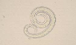
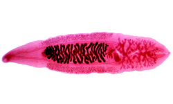
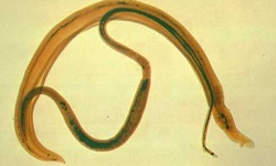
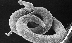
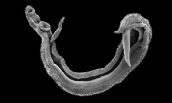
Image: unstained Fasciola hepatic adult (Source: CDC)
Laboratory diagnosis of Fasciola hepatica
The laboratory diagnosis of Fasciola hepatica starts with the collection of samples, followed by microscopy, serodiagnosis, and imaging methods.
Sample
Samples collected include:
stool
biopsy tissues (liver, skin, lung, heart, brain, eye, intestine)
blood (serum)
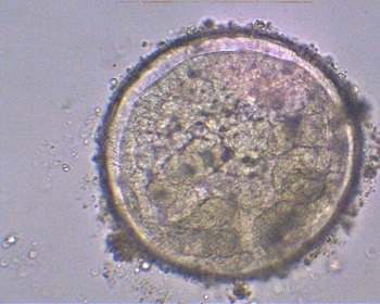
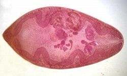
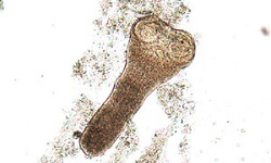
Image: Fasciola hepatic metacaercriae (Source: ar.javamem)
Microscopy
microscopic diagnosis of fascioliasis is commonly done by demonstration of Fasciola hepatica eggs
useful to determine chronic fascioliasis but not acute fascioliasis
in acute cases, eggs are scanty and intermittent
concentrations are done by sedimentation in 0.5% glycerinated saline, formalin-ether concentration method
cannot distinguish between Fasciola hepatica and Fasciola buski eggs
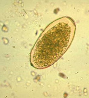
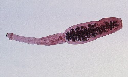
Image: Fasciola hepatic egg (Source: ruby.fgcu.edu)
Stool antigen detection
Enzyme-linked immunosorbent assay (ELISA) can be done in stool samples to detect Fasciola corpo-antigen for chronic infection
Serodiagnosis
Serodiagnostic methods can be done for early detection of liver fluke infection as these tests are able to demonstrate infection in the serum within 2- 4 weeks of infection – which is 4 to 8 weeks before eggs are released in the stool.
serodiagnosis for Fasciola hepatica can detect the infection early – during the pre-patent period which prevents liver damage due to chronic fascioliasis
useful to detect acute fascioliasis and ectopic fascioliasis as eggs are not passed in the faeces in such conditions
prevents pseudofascioliasis in which eggs are seen in the stool but is not due to active infection but due to ingestion of raw sheep or cattle liver containing Fasciola hepatica eggs
Numerous methods used for serodiagnosis include:
immunoelectrophoresis
indirect haemagglutination
indirect immunofluorescence
Western blot - 100% specificity when 12kDa, 17kDa, and 63kDa Fasciola hepatica antigen are used
enzyme-linked immunosorbent assay (ELISA) – has 95% sensitivity and
* uses excretory-secretory antigen of Fasciola hepatica
Intradermal Skin Test
The intradermal skin test involves the administration of fascioline into the intradermal layer of the patient’s skin. Fascioline is an extract of adult Fasciola, with a nitrogen content of 0.3ng/ml.
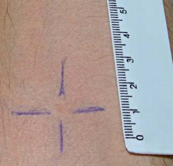
Image: Fasciola hepatic intradermal skin test (Source: CDC)
Molecular test
Important molecular tests of Fasciola hepatica diagnosis include:
DNA probes
PCR
Fasciola hepatica and Fasciola gigantica can be differentiated by the use of protein banding patterns after isoelectric focusing.
Imaging Methods
Ultrasonogram (USG) shows hypoechoic lesions in the liver made by migrating larvae, as well as adult Fasciola hepatica, flukes in the gallbladder or the bile duct
CT-scan
MRI
Treatment of Fasciola hepatica infection
Drug of choice for the treatment of Fasciola hepatica infection is bithionol and Praziquantel.
Prevention, Control of Fasciola hepatica
The control and prevention of Fasciola hepatica can be obtained by following steps:
avoid consumption of raw/undercooked sheep/cattle liver
thorough cleaning and decontamination of salad vegetables such as watercress and other water-grown greens
proper filtration and/or boiling of drinking water
immediate treatment of infections in sheep, cattle, and people
use of molluscicides to control snails
reduce contamination of water sources by animal faeces