Giardia intestinalis - Classification, History, Habitat, Morphology, Culture
Introduction of Giardia intestinalis
Giardia intestinalis, also known as G. lamblia or G. duodenalis inhabits the intestinal tract of humans. They are the only intestinal parasitic flagellate known to cause both endemic and epidemic diarrhea in humans.
Classification of Giardia intestinalis
Giardia intestinalis is classified:
Domain: Eukaryota
Phylum: Metamonada
Order: Diplomonadida
Family: Hexamitidae
Subfamily: Giardiinae
Genus: Giardia
Species: intestinalis
History of Giardia intestinalis
Histologically, in 1681, Antony von Leeuwenhoek first observed the flagellate when examining his own stool. Lambl coined the name Giardia lamblia in 1859 after observing it in human stool. Only in the 1970s, the flagellate was recognized as a parasite as protozoa were considered a commensal organism before this.
Habitat of Giardia intestinalis
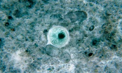
Giardia intestinalis inhabits the small intestine of humans and the trophozoites, cysts are found in the duodenum, jejunum, and upper ileum of the intestine.
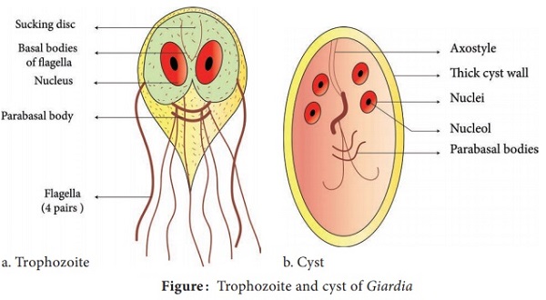
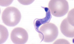
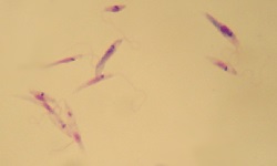
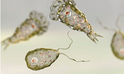
Fig: G. intestinalis morphology (Source: BrainKart)
Morphology of Giardia intestinalis
The Giardia intestinalis parasite occurs in two stages- trophozoites and cysts
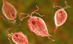
Trophozoite
this morphological form of Giardia intestinalis is 9μm – 21 μm in length and 5μm- 15μm in breadth
trophozoites are pear-shaped, bilateral bodes with a broad rounded anterior end and a tapering posterior end
since their dorsal surface is convex and ventral surface is concave, they appear sickle-shaped when viewed from a lateral view
it bears two median bodies, two axonemes, four pairs of flagella (two pairs lateral, one pair ventral, and one pair caudal), two nuclei
each of the two nuclei contains a larger central karyosome
the axostyles run diagonally across the cytoplasm which is uniform and finely granular
a bilobed adhesive disc is present on one-third to one-half of the ventral surface
* These discs are rigid structures that help in attachment to the host cells
giardin is a tubulin intermediate filament that is a part of the cytoplasmic extension
median bodies are a pair of large, curved, and transverse bodies present behind the adhesive discs
Although the function of the median bodies is unclear, it is believed to help in energy metabolism
motility takes place with the help of flagella, showing the typical “falling leaf motility”
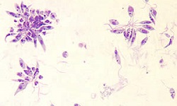
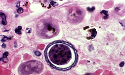
Cyst
cyst is the infective form of G. lamblia
oval or ellipsoidal in shape, measuring 8μm-12μm
a thick, finely granular cytoplasm cyst wall surrounds the cyst and is separated from the cyst wall by a thin, clear space
it contains four nuclei, axostyle, and median bodies
if stained with iodine, the cysts take brown color
Culture of Giardia intestinalis
Giardia intestinalis can be cultured in media as well as laboratory animals.
In media
Giardia intestinalis can be grown axenically in Diamond’s medium, containing Entamoeba histolytica. It is mostly used for research purposes and rarely for routine diagnosis
In animals
Giardia intestinalis is inoculated into laboratory mice to study the host immune response and pathogenesis of malabsorption in giardiasis.