Trypanosoma cruzi - Introduction, History, Habitat, Morphology, Culture
Introduction of Trypanosoma cruzi
Trypanosoma cruzi are flagellated protozoa causing a zoonotic disease called Chagas’ disease or South American trypanosomiasis. They commonly occur in South American countries. This is the only Trypanosoma parasite that can be transmitted by feces of the invertebrate vector while other Trypanosoma species are transmitted by saliva.
History of Trypanosoma cruzi
Historically, Brazilian physician Carlos R.J. Chagas first discovered Trypanosoma cruzi in 1901 in the intestine of the vector i.e. reduviid bug. This parasite was later found to cause disease in humans.
Habitat of Trypanosoma cruzi
In humans, Trypanosoma cruzi exists in two morphological forms – amastigote and trypomastigote.
The amastigote forms are intracellular and are found inside the reticuloendothelial cells, mononuclear phagocytes, and muscles.
On the other hand, trypomastigotes habitats the peripheral blood.
Morphology of Trypanosoma cruzi
The occurrence of different morphological forms of Trypanosoma cruzi depends on the host where the parasite is infesting.
Vertebrate forms
In vertebrate hosts, Trypanosoma cruzi occurs in two morphological forms- Amastigote and non-multiplying trypomastigotes.
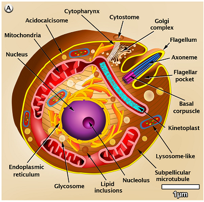
Figure: Trypanosoma cruzi - amastigote morphology (Source: journals.plos)
Amastigote
Trypanosoma cruzi in amastigote forms multiply in humans during this stage only
this is a non-flagellated and intracellular replicative form
round or oval body
body measures 2μm - 4μm in diameter
it has a nucleus, kinetoplast, and axoneme but lacks flagellum
also called leishmanial form as Trypanosoma cruzi amastigotes are similar to amastigotes of Leishmania spp.
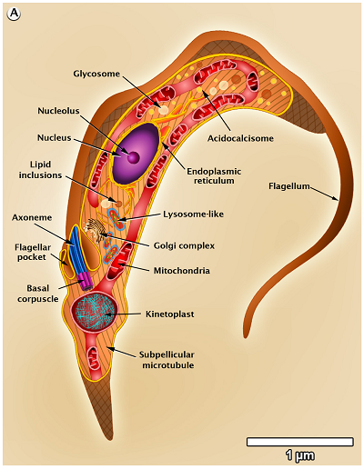
Figure: Trypanosoma cruzi - trypomastigote morphology (Source: journals.plos)
Non-multiplying form/trypomastigote
Non-multiplying form/trypomastigote of Trypanosoma cruzi are non-multiplying forms
measures 2μm - 4μm in breadth
found in the peripheral blood of mammal hosts, including the man
the body is C-shaped and slender with a wedge-shaped posterior end
the nucleus is centrally placed while the large oval-to-round kinetoplast is situated at the posterior end
a flagellum, which originates from the kinetosome and transverses on the surface of the trypomastigote as a narrow undulating membrane
Insect form
The insect form of Trypanosoma cruzi occurs in the invertebrate host
exists in two forms- epimastigotes and multiplying trypomastigote
these forms are found in the end part of insect vector (reduviid bug) digestive and urinary tract
also found in culture
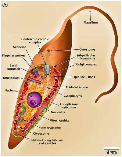
Figure: Trypanosoma cruzi - epimastigotes morphology (Source: journals.plos)
Culture of Trypanosoma cruzi
The culture of Trypanosoma cruzi can be done in media and laboratory animals.
In media
the metacyclic trypomastigotes forms of Trypanosoma cruzi are grown in the NNN media and the LMC media
can also be grown in Graces’ insect tissue culture medium supplemented with 10% newborn calf serum maintained at 6.6 pH
Laboratory animals
Trypanosoma cruzi can be cultured in mice
frequently used to isolate the parasite from clinical samples