Hymenolepis nana - Classification, History, Morphology, Epidemiology, Reservoir, Transmission
Introduction of Hymenolepis nana
Hymenolepis nana is the cause of hymenolepiasis in humans, the other species being H. diminuta. These tapeworms have broader proglottids than longer proglottids. The number of testes varies from 1 to 4 in number and genital pores are always present. Also, the unilateral uterus appears saclike under a microscope.
Hymenolepis nana is a common parasite in man which also is the smallest cestode infecting humans. Thus, it is also called the dwarf tapeworm (nana = dwarf). It is the only cestode that does not require an intermediate host.
Classification of Hymenolepis nana
Kingdom: Animalia
Phylum: Platyhelminthes
Class: Cestoda
Order: Cyclophyllidea
Family: Hymenolepididae
Genus: Hymenolepis
Species: H. nana
History of Hymenolepis nana
The tapeworm was first discovered in the small intestine by Bilharz in 1851.
Habitat of Hymenolepis nana
The adult Hymenolepis nana is present in the ileal portion of the small intestine of man and other mammals such as mice, and rats.
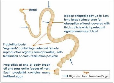
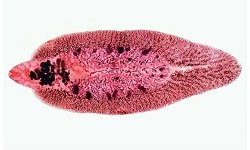
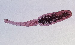
Image: Hymenolepis nana adult morphology (Source: Invertebrate Diversity)
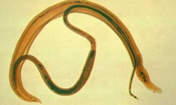
Morphology of Hymenolepis nana
Adult form
thread-like tapeworm
small – measures 10 cm to 40 cm in length
consists of a head (scolex), neck, and body (strobila)
lives for nearly two weeks
nearly 1000 to 5000 adults tapeworms may infect a man spontaneously
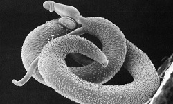
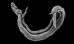
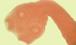
Scolex
globular with four suckers and a short rostellum
has 20 to 40 hooklets
the retractable rostellum always remains invaginated at the apex of the organ
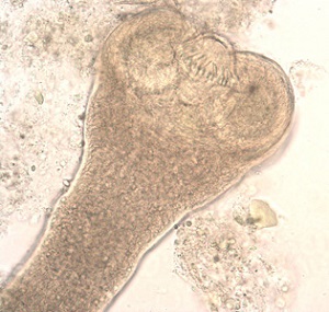
Image: Hymenolepis nana scolex (Source: CDC)
Neck
long and is situated posterior to the scolex
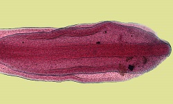
Strobila
contains nearly 200 segments or proglottids
each mature segment measures 0.3mm in length and 0.9mm in breadth
as segments move away from the scolex, it develops and matures
genital pores are situated on the same side along the margin
gravid proglottids contain fertilized eggs
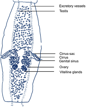
Image: Hymenolepis nana proglottid (Source: Springer Link)
Infective form
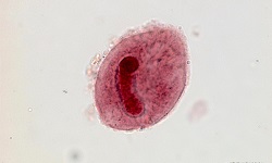
Egg
colorless and oval in shape
size in diameter ranges from 30μm to 45 μm
have two membranes separated by a clear space in between filled with yolk granules
outer membranes – thin, colorless
inner membranes (embryophore) – encloses an oncosphere with three pairs of hooklets
the inner membrane has two poles from where 4 to 8 thread-like polar filaments emerge and fill the space between the two membranes
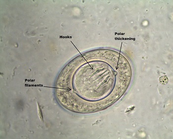
Image: Hymenolepis nana egg (Source: ASM)
Epidemiology of Hymenolepis nana
Hymenolepis nana is cosmopolitan in distribution and is the most common cestode-causing infection.
The tapeworm is highly prevalent in South Africa, South Europe, Middle East Asia, and Latin America- mostly in rural areas.
Reservoir, Source of Hymenolepis nana
Feces from infected humans are the chief source of infection.
Man is the main reservoir while the hexacanth eggs are the infectious form of Hymenolepis nana.
Transmission of Hymenolepis nana
The transmission of Hymenolepis nana occurs through the following route:
fecal-oral route by ingestion of eggs from contaminated food and water
ingestion of eggs from contaminated hands due to bad personal hygiene
consumption of food contaminated with fleas harboring cysticercoid larvae
Complications of Hymenolepis nana
Hymenolepis nana infections become rarely complicated. These include diarrhea and behavioral disturbances.
Prognosis of Hymenolepis nana
Even without treatment, the prognosis is excellent.