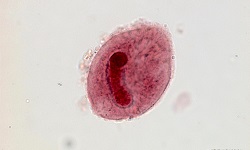
Hymenolepis nana - Life Cycle, Pathogenesis, Pathology, Host Immunity, Clinical Manifestation, Laboratory Diagnosis
Life Cycle of Hymenolepis nana
Hymenolepis nana is the only cestode that does not require any intermediate host and the life cycle is completed in a single host. The definitive host includes humans, a rat, and a mouse.
humans become infected after ingestion of Hymenolepis nana eggs
once in the small intestine, the embryo (oncosphere) is released from the egg
the oncosphere penetrates the small intestine and within 5 days, it develops into a small larval stage (cysticercoid)
after fully maturing, the larva travels back to the small intestinal lumen where it attaches itself to the wall of the intestine with the help of suckers
within a month of infection, the tapeworm rapidly develops into an adult and the gravid proglottid breaks off and disintegrates to release fertilized eggs
these eggs are passed out along with the host feces which is the source of infection for new hosts (direct infection)
in some cases, the eggs hatch in the small intestine lumen of the original host to release embryos in the process called internal autoinfection
autoinfection is responsible for the tremendous increase in the number of tapeworms in the intestine
external autoinfection may also occur- which occurs when a person ingests eggs dispersed from their own body via the fecal-oral route and is mostly due to a lack of personal hygiene
moreover, external autoinfection is responsible for the persistence of Hymenolepis nana infection
by the process of direct infection, internal or external autoinfection, the life cycle of Hymenolepis nana continues
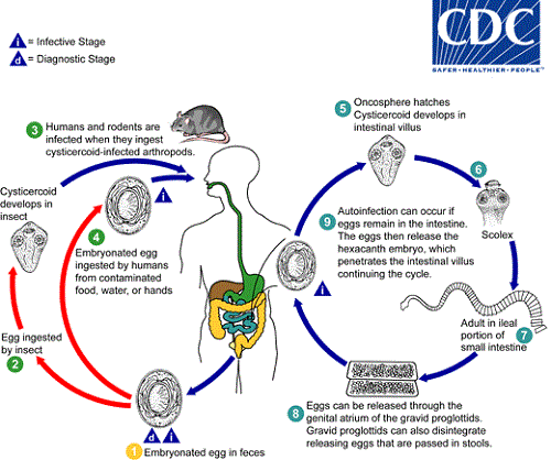
Image: Hymenolepis nana life-cycle (Source: CDC)
Pathogenesis, Pathology of Hymenolepis nana
Infection by cysticercoid larvae of Hymenolepis nana in the intestine shows little to no pathology. However, in heavy cases of infection, pathological changes include:
mechanical irritation of the intestine
release of toxic metabolites results in various allergic manifestations including anal and nasal pruritus
Host Immunity of Hymenolepis nana
If Hymenolepis nana infection is acquired by direct cycle, it evokes a strong humoral and cellular response from the host body. The host immunity provides protection from subsequent infection by the same tapeworm.
But the immune system is unable to remove adult worms that have attached and invaded the gut wall.
Clinical Manifestations of Hymenolepis nana
Hymenolepiasis is more common in children (age 4-10 years) than in adults, mostly due to a lack of proper regular hygiene.
the incubation period is between 15 days to 39 days
mostly asymptomatic
in cases of heavy infections, symptoms include abdominal irritation, diarrhea, abdominal pain, anal pruritis, nasal pruritis, and sleep disorders
in rare cases, symptoms include nausea, vomiting, anorexia
Laboratory diagnosis of Hymenolepis nana
Sample
Stool
Microscopy
Direct wet mount microscopy
the presence of Hymenolepis nana eggs in stool samples confirms the infection
the wet mount is prepared by mixing the sample with saline, iodine, or lactophenol cotton blue (LPCB)
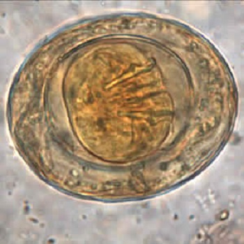
Image: Hymenolepis nana egg (Source: CDC)
Examination of a stool after concentration
formalin-ethyl acetate and salt floatation concentration methods are used to concentrate the stool so that the parasites have increased yield
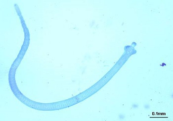
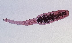
Image: Hymenolepis nana adult (Source: Medical Pictures Info)
Treatment of Hymenolepis nana
Drugs of choice include praziquantel, niclosamide, and paromomycin. Due to the presence of a large number of tapeworms due to autoinfection and the presence of cysticerci, repeated treatment is required for a period of two weeks.
Prevention, Control of Hymenolepis nana
Individual prophylaxis
improved personal hygiene such as proper washing of hands with soap after defecation and before eating food
treatment of water before drinking such as boiling, filtering the water used in a 0.22-micrometer membrane, iodination with tetracycline hydro per iodide
drinking bottled water while traveling to areas endemic to hymenolepiasis as chlorine treatment cannot kill the eggs
if salad is to be consumed, treat the vegetables with acetic acid or vinegar for 15 minutes
not performing sexual acts that involve fecal-oral contact
Community prophylaxis
improvement of the water management system to avoid fecal contamination
improvement of sanitation by installing latrines for proper disposal of human feces