Balantidium coli - Introduction, Classification, History, Habitat, Morphology, Culture
Introduction of Balantidium coli
Balantidium coli are exclusive parasites of the vertebrate and invertebrate hosts inhabiting their digestive tract. They multiply by binary fission while their sexual cycle takes place by conjugation.
Balantidium coli is the only species in the genus Balantidium which is pathogenic to humans. Also, it is the largest human protozoa parasite.
Balantidium coli are oval in shape with conspicuous cytostome, contractive vacuoles, two nuclei (large bean-shaped macronucleus and minute micronucleus), and cilia over their body.
Classification of Balantidium coli
The classification of Balantidium coli is as follows
Kingdom: Chromista
Superphylum: Alveolata
Phylum: Ciliophora
Class: Litostomatea
Order: Vestibuliferida
Family: Balantidiidae
Genus: Balantidium
Species: B. coli
History of Balantidium coli
The human protozoa parasite was first described in 1857 by Malmsten in the feces of a man suffering from dysentery and named it Paramecium coli. It was later named Balantidium coli by Stein in 1863.
Habitat of Balantidium coli
Balantidium coli inhabits the intestine of humans, pigs, and monkeys.
Morphology of Balantidium coli
The morphological stages of Balantidium coli include trophozoite and cyst.
Trophozoite
trophozoites are the invasive form of Balantidium coli
size varies from 30 μm to 300 μm in length and 30 μm to 100 μm in breadth
oval-shaped with a slightly pointed anterior end while the posterior end is rounded
Balantidium coli motility takes place with the help of rows of tiny cilia which covers the entire surface of the body
conspicuous V-shaped mouth called cytostome present at the anterior end
cytostome leads to the cytopharynx which is a funnel-shaped guller extending up to one-third of the body
a less prominent pore called cytostome is present at the posterior end of the parasite
cytostome function is to evacuate the contents of food molecules
two nuclei (large bean-shaped macronucleus and minute micronucleus) are present
micronucleus lies on the concavity of the macronucleus
numerous food vacuoles and two contractile vacuoles are also present
the trophozoite of Balantidium coli feeds on bacteria, erythrocytes, and fat droplets
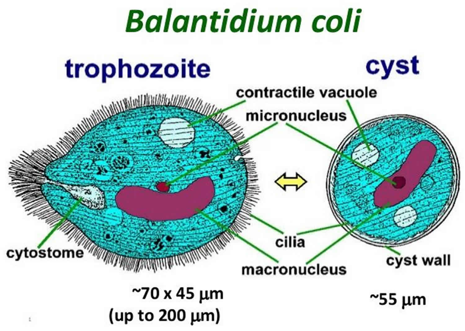
Figure: Balantidium coli morphology - trophozoite, cyst (Source: Health Jade)
Cyst
Balantidium coli cysts are resistant form as well as the infective form
round in shape
cysts are smaller than trophozoites
measures 40 μm to 60 μm in diameter
a cyst is surrounded by a cyst wall which is thick and transparent
like trophozoite form, the Balantidium coli cyst also contains two nuclei (large bean-shaped macronucleus and minute micronucleus) and the micronucleus lies on the concavity of the macronucleus
granular cytoplasm has a refractile body and numerous food vacuoles
cilia may be present in younger cysts but are absent in mature ones
Culture of Balantidium coli
In media
Balantidium coli can be grown in Robinson’s medium. Culture media used for in-vitro culture of Entamoeba histolytica.
NIH Polyxenic culture
created by Boeck and Drbohlav in 1927
contains the egg-serum medium with Lock’s medium and bacterial flora (polyxenic or polybacterial culture) which will provide nourishment for the growing parasites
other cultures such as Balamuths, Nelsons, or Robertsons’ media can also be used to isolate the parasite from the stool
Axenic Culture
this bacteria-free culture was first created by Diamond in 1961
useful for:
studying the pathogenicity of protozoa
test anti-parasite drugs in vitro
study immunological property
prepare axenic amoebic antigens for immunodiagnosis