Leishmania braziliensis, Leishmania mexicana - Clinical manifestations, Epidemiology, Reservoir, Source, Transmission
Clinical manifestations of Leishmania braziliensis, Leishmania mexicana
Leishmania braziliensis Complex
Leishmania braziliensis causes the following clinical syndromes
Espundia
espundia most severe and destructive form than the oriental sore
mucocutaneous lesions are chronic, resistant to treatment
single or multiple ulcers are present in the lower extremities
the ulcers might not heal completely or spontaneously
if metastasis occurs, lesions may appear in the mouth, nasopharynx even months to years after the occurrence of the primary lesion
the perforation and severe destruction of the nasal septum through the soft palate or skin of the nose may result in the collapse of the nose, the condition termed tapir nose
in advanced cases of espundia, soft tissues such as the upper lip, tongue, and buccal, pharyngeal, and laryngeal mucosa may occur
other symptoms include fever, anemia, and weight loss
death may occur due to secondary infections, pneumonia, or respiratory obstruction
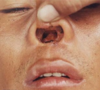
Image: Tapir nose in Espundia (Source: ResearchGate)
Pian bois
pain bois is also known as forest yawns
caused by Leishmania braziliensis guyanensis
single or multiple ulcers, which are dry, painless, and persistent appear all over the body
Uta
uta is caused by Leishmania braziliensis peruviana
ulcers, ranging from one to several in number, may be present on the face
the nasopharynx is not affected
lesions heal spontaneously within 3 months to a year
Non-healing ulcerate
non-healing ulcerate caused by Leishmania braziliensis panamensis
a single non-healing ulcer is present
lymph nodes are present
Leishmania mexicana Complex
Leishmania mexicana causes the following clinical syndromes
Chiclero ulcerate
chiclero ulcerate is also known as bay sore
caused by Leishmania mexicana mexicana
ulcers appear on the hand and head and are persistent for years
although these ulcers heal spontaneously, they can entirely destroy the pinna of the ear
Indolent nodular lesion
indolent nodular lesion is caused by Leishmania mexicana venezuelensis
Cutaneous single sore type
cutaneous single sore type is caused by Leishmania mexicana amazonensis
can cause diffuse cutaneous leishmaniasis
Diffuse cutaneous leishmaniasis
usually, diffuse cutaneous leishmaniasis condition arises in hosts with poor immune response and is allergic
the disease begins as a localized papule, lacks ulceration, and satellite lesions may appear around the papule in the skin
the amastigotes can metastasize to the face and other extremities
as this is a chronic infection, the syndromes may last 20 or more years
the Leishmania skin test is negative in these cases
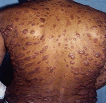
Image: Diffuse cutaneous leishmaniasis (Source: ResearchGate)
Epidemiology of Leishmania braziliensis, Leishmania mexicana
Epidemiological studies have confirmed that the new world cutaneous and mucocutaneous leishmaniasis are endemic to South and Central America- mostly in Bolivia, Brazil, and other Latin American countries.
Reservoir, Source of Leishmania braziliensis, Leishmania mexicana
Except for the condition uta, New World cutaneous and mucocutaneous leishmaniasis are zoonotic in nature. Mammals such as dogs, sloths, anteaters, and rats are reservoirs, sources of Leishmania braziliensis Complex and Leishmania mexicana Complex.
Transmission of Leishmania braziliensis, Leishmania mexicana
Transmission of mucocutaneous and cutaneous leishmaniasis, caused by Leishmania braziliensis, Leishmania Mexicana, occurs mostly by the bite of the sandfly vector. The infection is seen most commonly in individuals working at the edge of the forests and in rural areas.
They are less frequently transmitted by ticks, direct man-to-man transmission, and auto-infection.