Leishmania braziliensis, Leishmania mexicana - History, Habitat, Culture, Morphology
Introduction of Leishmania braziliensis, Leishmania mexicana
Leishmania braziliensis Complex and Leishmania mexicana Complex cause New World cutaneous and mucocutaneous leishmaniasis. The lesions may vary from a single localized ulcer in the skin to spreading lesions such as espundia.
Depending upon their geographical location, mucocutaneous leishmaniasis has several names- espundia, pian bois, bubas braziliana, uta, chiclero ulcer, etc.
History of Leishmania braziliensis, Leishmania mexicana
Historically, in 1909, Lindenberg and Paranhos were the first ones to describe amastigotes in ulcers of skin from a man in Brazil. Carini in 1911 reported observing amastigotes in ulcers and nasopharyngeal mucous membranes. The same year, in 1911, Vianna named the species Leishmania braziliensis.
Habitat of Leishmania braziliensis, Leishmania mexicana
Leishmania braziliensis are intracellular parasites and habitats inside the macrophages of the skin as well as the mucous membranes of the nose and buccal cavity.
Unlike Leishmania donovani, they are absent from peripheral blood and internal organs.
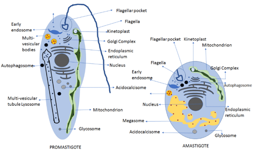
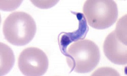
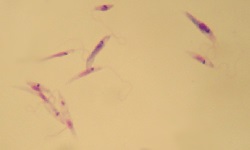
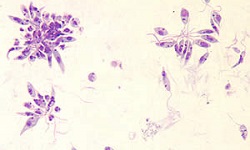
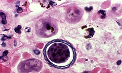
Figure: Leishmania braziliensis Complex and Leishmania mexicana Complex morphology (Source: ResearchGate)
Morphology of Leishmania braziliensis, Leishmania mexicana
The morphology of Leishmania braziliensis Complex and Leishmania mexicana Complex is similar to that of Leishmania donovani. The protozoa parasites exist in two forms- amastigote and promastigote form.
Amastigote form
Amastigote form is found in humans and other mammal hosts
small, round to oval bodies
each amastigote form measures 2-3μm in length
resides inside monocytes, endothelial cells, polymorphonuclear leucocytes of the host
the nucleus is large and lies at a right angle to the kinetoplast which is slender, rod-shaped
axoneme arises from the kinetoplast and extends to the margin of the amastigote
vacuole, which is not stained lies alongside the axoneme
stained well in Giemsa and Wright stain
in Giemsa-stained preparations, the nucleus, and kinetoplast, are stained red
Promastigote
promastigote occur naturally in the digestive tract of sand fly (vector)
mature promastigotes are long, slender, and spindle-shaped
measures 15 μm to 25 μm in length and 1,5 μm to 3.5 μm in breadth
a single nucleus is centrally located
kinetoplast located transversally near the anterior end
the presence of a single flagellum measuring 15μm -28 μm
Leishman stain stains the cytoplasm blue, the nucleus pink, and the kinetoplast bright red
Culture of Leishmania braziliensis, Leishmania mexicana
Leishmania braziliensis, Leishmania mexicana can be cultured in artificial media as well as in laboratory animals.
In media
The Leishmania braziliensis Complex and Leishmania mexicana Complex can be cultured in biphasic media, liquid media, or laboratory animals. They are slow growers and require more incubation time.
Biphasic media
consists of two parts of salt agar and one part defibrinated rabbit blood
Novy and McNeal (1904) and Nicolle (1908) (NNN) medium was the first biphasic medium for the culture of Leishmania braziliensis Complex and Leishmania mexicana Complex
Liquid media
includes Schneider’s, Grace’s, and Mituhasi-Maramorosh media- which are insect cell culture media
liquid media do not contain any blood
usually used for the preparation of a large volume of promastigotes
Schneider’s medium which contains 20% fetal calf serum (FCS) is more sensitive to culture than the NNN biphasic medium
Laboratory animals
Chinese hamsters and golden hamsters are routinely used in the culture and diagnosis of Leishmania braziliensis Complex and Leishmania mexicana Complex.