Leishmania braziliensis, Leishmania mexicana- Laboratory diagnosis, Treatment, Prevention, Control
Laboratory diagnosis of Leishmania braziliensis, Leishmania mexicana
The laboratory diagnosis of Leishmania braziliensis, Leishmania mexicana begins with the collection of samples.
Specimen
The lab diagnosis of Leishmania braziliensis Complex and Leishmania mexicana Complex includes:
skin- biopsy
ulcer edge aspirates
nodule aspirates
Microscopy
stains such as Leishman stain, Giemsa stain, Brown-Hopps stain, or Wright stain can be used for microscopy
presence of amastigotes confrims the diagnosis
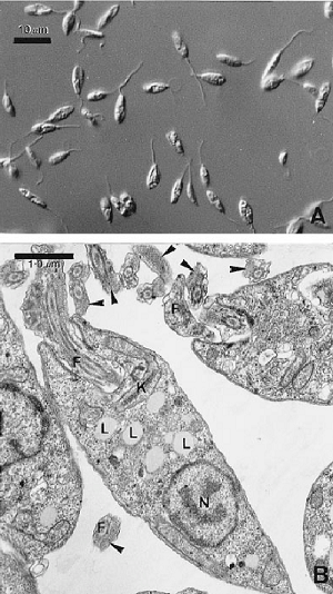
Image: Morphology of Leishmania braziliensis promastigotes - K, kinetoplast; N, nucleus; F, flagellum; L, lipid droplets (Source: ResearchGate)
Culture
Leishmania braziliensis and Leishmania mexicana can be cultured in the laboratory both in lab animals and in-vitro culture.
In-vitro culture
media used includes NNN, Schneider Drosophila medium, or any biphasic medium
samples are inoculated into the water of condensation or into the fluid of a liquid medium
incubated at 22° C - 26° C for 1-4 weeks
at the end of each week, a drop of culture is microscopically examined for promastigotes
motile promastigotes can be observed in positive cultures
has a sensitivity of 50% - 75%
Leishmania mexicana complex grows well in culture while Leishmania braziliensis braziliensis grow poorly or do not grow at all in media cultures
Animal inoculation
laboratory animals such as Chinese and golden hamsters are inoculated intraperitoneally
skin biopsy, lesion aspirations are stained and visualized under a microscope
Leishmania mexicana complex grows fast and produces lesions on the skin while Leishmania braziliensis braziliensis grows slowly and takes time to produce a pathological lesion
Serodiagnosis
The serodiagnosis of Leishmania braziliensis and Leishmania mexicana includes the following methods.
Indirect Immuno-fluorescent (IFA)
uses fixed amastigotes as antigen
positive in 89% - 95% of cases
used to evaluate the response if the patient is treated with chemotherapy as IFA titers fall after successful chemotherapy
Enzyme-Linked Immunosorbent Assay (ELISA)
uses fixed amastigotes as antigen
sensitivity of up to 85%
Leishmania skin test (Montenegro test)
Leishmania braziliensis and Leishmania mexicana can also be diagnosed by the Leishmania skin test (Montenegro test).
delayed hypersensitivity test and is a helpful test
positive skin test results are also seen in cutaneous and mucocutaneous leishmaniasis
negative in cases of diffuse cutaneous leishmaniasis
0.2 ml of Leishmania antigen is injected intradermally and read after 48-74 hours
induration and erythema of 5mm diameter or larger are seen in positive cases
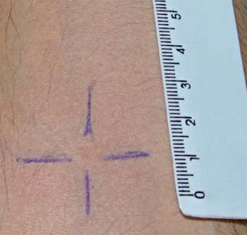
Image: Leishmania skin test (Source: ResearchGate)
Molecular test
Following molecular methods are used for the diagnosis of Leishmania braziliensis, and Leishmania mexicana.
DNA probes
PCR
Treatment of Leishmania braziliensis, Leishmania mexicana
Drugs used for the treatment of Leishmania braziliensis, and Leishmania mexicana infection include:
Pentavalent antimonials
megalumine antimonate
sodium stibugluconate solution
Pentamidine
pentamidine isethionate
pentamidine dimethane
Amphotericin B
Miltefosine
Interferon
Prevention, Control of Leishmania braziliensis, Leishmania mexicana
Prevention, Control of Leishmania braziliensis, and Leishmania mexicana can be done by:
reduction of sand-fly population by using insecticides
evade contact with potential animal vectors such as dogs, foxes, and rodents
use of bed nets, window nets, or insect repellents