Leishmania braziliensis, Leishmania mexicana - Life Cycle, Pathogenesis, Pathology, Host Immunity
Life Cycle of Leishmania braziliensis, Leishmania mexicana
The life cycle of Leishmania braziliensis Complex and Leishmania mexicana Complex is completed in two different hosts:
Man or other mammals- the amastigote form
Sandfly of genus Phlebotomus and Lutzomyia – the promastigote form
These mammals include dogs, rodents, foxes, etc.
Leishmania braziliensis Complex and Leishmania mexicana Complex are transmitted to man and other vertebrates by the bite of a blood-sucking female sandfly
During the blood meal, the promastigotes are transmitted from the saliva of the vector to the host’s skin
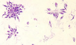
the promastigotes are then immediately phagocytosed by the host macrophages where they transform into amastigotes
the newly formed amastigotes undergo binary fission to produce a large number of amastigotes which continue to divide inside the enlarged macrophages
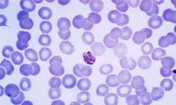
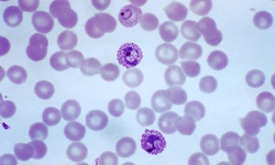
the infected macrophages rupture to release a large number of amastigotes (around 200 in number) into the host body circulation
the parasites then reside in the reticuloendothelial cells and lymphatic tissues of the skin but do not invade the internal organs or peripheral blood
if the infected mammal host (including man) is bitten by a female sand-fly during a blood meal, the free amastigotes are also taken up with the host blood
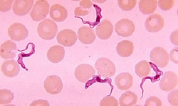
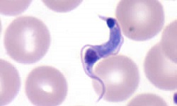
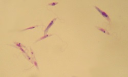
in the midgut of the sand-fly, within 72 hours, the amastigotes are transformed into promastigotes
the morphological transformation takes place through a series of flagellated intermediate promastigote forms to flagellated promastigotes
the promastigotes undergo binary fission, produce a large number of parasites, and completely fill the lumen of the gut
after 6-9 days, the promastigotes travel to the sandfly's pharynx and buccal cavity from the mid-gut
bite from the infected sand-fly to mammals (including humans) during its blood meal transmits the infection and the life cycle of Leishmania braziliensis Complex and Leishmania mexicana Complex is continued
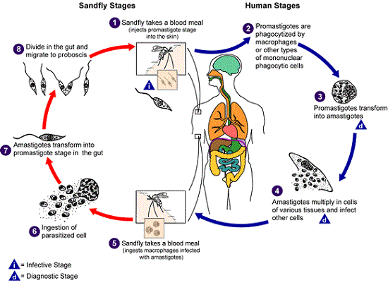
Figure: Leishmania braziliensis Complex and Leishmania mexicana Complex life cycle (Source: CDC)
Pathogenesis of Leishmania braziliensis, Leishmania mexicana
A bite from a female sandfly during a blood meal deposits the promastigotes, present in the vector’s saliva/mouthparts, on the surface of the skin. The newly introduced promastigotes are phagocytosed by host macrophages where they transform into amastigotes.
The key pathological lesions of Leishmania braziliensis Complex and Leishmania mexicana Complex infection are papules and ulcers, which are the common forms of mucocutaneous leishmaniasis.
Pathology of Leishmania braziliensis, Leishmania mexicana
The pathology of Leishmania braziliensis, Leishmania mexicana are as follows:
Espundia
the incubation period for oriental sore may range from 2 months to 4 months
espundia is a typical ulcerative lesion that occurs on the skin
the syndrome begins as a single, red, pruritic papule at the site of the bite i.e. at the site of inoculation
lesions may expand rapidly and forms large, long-lasting ulcers with weeping surfaces and distinct margins
as the size of the papule increases, it becomes crusted and eventually ulcerates and this ulcer is called an oriental sore
the oriental sore is circular, shallow, and may measure around 2 cm with a base formed by granulation tissue and erythematous elevated margin
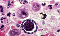
the base of the ulcer may contain the amastigote forms of the parasite
as the disease progresses, the ulcer produces serous or seropurulent discharges which may result in the formation of scales or crusts
the ulcer may also cause secondary infections such as streptococcal and staphylococcal infections
regional lymphadenopathy is mostly unusual
in some cases, the ulcer has lasted for 1 year to 2 years, and healing leaves behind small, flat, and depigmented scar
in 5% of espundia cases, mucosal metastasis may occur by direct extension or through blood and invade mucous membranes in the mouth, nose, pharynx, and larynx either
Ulcer
primary ulcers occur mostly in the hand and foot
are round in shape with raised bodies and a granulating base covered by the exudate
a chain of nodules may appear along the line of lymphatics due to lymphatics draining these ulcers
mucosal leishmaniasis ulcers are fungative, indurated, invade and destroy superficial tissues and cartilages of the nose and larynx
under a microscope, the edge of the ulcer and the serum exudate reveals the presence of amastigotes
Host Immunity of Leishmania braziliensis, Leishmania mexicana
Host Immunity of Leishmania braziliensis, Leishmania mexicana involves cell-mediated immunity (CMI) while the role of humoral immunity is unclear.
mucocutaneous leishmaniasis involves the development of marked cell-mediated hypersensitivity reaction
as the cell-mediated immunity (CMI) is suppressed, amastigotes are freely able to multiply and spread in the skin
circulating antibodies are seen in the patient's serum