Leishmania donovani - Laboratory Diagnosis
Laboratory Diagnosis of Leishmania donovani
The laboratory diagnosis of Leishmania donovani includes:
Specimen
biopsy samples from the spleen, bone marrow, lymph node, liver, and peripheral blood smear
spleen aspiration – 98% sensitivity but risky as seen rupture can be fatal
bone marrow aspiration (collected from the sternum, iliac crest) – 85% sensitivity
lymph node aspiration – 60% sensitivity (useful for African leishmaniasis but not for Indian leishmaniasis)
peripheral blood smears – low sensitivity
Microscopy
Leishmania donovani amastigotes, also known as LD bodies) can be demonstrated in biopsy smears
stains such as Leishman stain, Giemsa stain, Brown-Hopps stain, or Wright stain can be used
LD bodies can be found within macrophages as well as freely in the host circulatory system which had recently escaped after rupturing from macrophages
observation of LD bodies in spleen aspiration, bone marrow aspiration, and lymph node aspiration also confirms infection by Leishmania donovani
in cases of stained peripheral blood smears, LD bodies can be microscopically demonstrated inside neutrophils and circulating monocytes
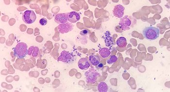
Image: L. donovani microscopy in case of Visceral Leishmaniasis (Source: Cureus)
Culture
Leishmania donovani can be cultured in laboratory animals as well as in-vitro culture.
In-vitro culture
media used includes NNN, Schneider Drosophila medium or any biphasic medium
tissue samples and aspirates are inoculated into the water of condensation or into the fluid of a liquid medium
incubated at 22° C - 26° C for 1-4 weeks
at the end of each week, a drop of culture is microscopically examined for promastigotes
motile promastigotes can be observed in positive cultures
has a sensitivity of 75%
Laboratory animal inoculation
laboratory animals such as Chinese and golden hamsters are inoculated intraperitoneally by Leishmania donovani culture
if the animal dies, its spleen smear is stained and visualized under a microscope
if the animal lives, they are killed after six months to visualize its stained spleen smear under a microscope
Serodiagnosis
Serodiagnosis is mostly useful to diagnose the early stages of visceral leishmaniasis caused by Leishmania donovani.
Complement Fixation Test (CFT)
Complement Fixation Test (CFT) is used to detect serum antibodies in cases of visceral leishmaniasis
Mycobacterium tuberculosis antigen WKK (Witebsky Klingenstein, Kuhn) and an antigen from Kedrowsky acid-fast bacillus are used
Indirect Immuno-fluorescent (IFA)
Indirect Immuno-fluorescent (IFA) uses cultured Leishmania donovani promastigotes as antigen
the disadvantage includes cross-reactivity with sera from leprosy, schistosomiasis, malaria, cutaneous leishmaniasis, and Chagas disease
Enzyme-Linked Immunosorbent Assay (ELISA)
Enzyme-Linked Immunosorbent Assay (ELISA) uses cultured Leishmania donovani promastigotes as antigen
the disadvantage includes cross-reactivity with sera from leprosy, schistosomiasis, malaria, cutaneous leishmaniasis, and Chagas disease
Direct Agglutination test (DAT)
Direct Agglutination test (DAT) uses trypsin-treated Coomasie blue-stained promastigotes as antigen
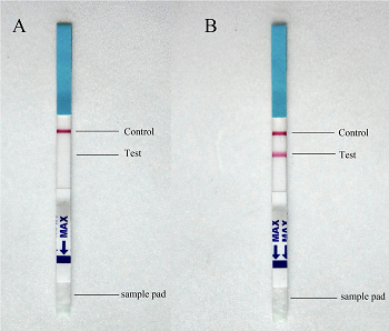
Image: Immunochromatographic strip test (Source: PLOS)
Strip test
Immunochromatographic strip test employs Leishmania rK39 antigen in the diagnosis of visceral leishmaniasis
Aldehyde test of Napier
Aldehyde test of Napier is done by adding a drop of 40% formalin to 1ml to 2 ml of serum in a test tube
within 2 to 20 minutes, if jellification occurs, changing the serum to a milky white opacity like of a boiled egg, the aldehyde test of Napier is positive
negative in cases of cutaneous leishmaniasis
disadvantages include cross-reactivity with cases of African trypanosomiasis, liver cirrhosis, schistosomiasis japonica, and multiple myeloma
Antimony test
Upon addition of urea stibamine solution into the patient's serum, antimony test results in the formation of a white flocculent precipitate in positive cases of leishmaniasis caused by Leishmania donovani.
Molecular test
The molecular tests used for diagnosis of Leishmania donovani are:
DNA probes
PCR
Leishmania skin test (Montenegro test)
Leishmania skin test (Montenegro test) delayed hypersensitivity test
gives negative results in active Leishmania donovani infections
indicates prior exposure in positive cases
positive skin test results are seen only 6 to 8 weeks after the cure
0.2 ml of Leishmania antigen is injected intradermally and read after 48-74 hours
induration and erythema of 5mm diameter or larger are seen in positive cases
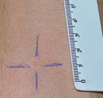
Image: Leishmania skin test (Source: ResearchGate)