Leshmania donovani - Classification, History, Habitat, Morphology, Culture
Introduction of Leishmania donovani
Leishmania donovani is protozoa causing the zoonotic infection called leishmaniasis. They are transmitted by the bite of an infected female sandfly during a blood meal to humans and other mammals. In humans, the incidental hosts of the intracellular parasite, infect the mononuclear phagocytes.
Leishmania donovani causes leishmaniasis which is also known as Dum-Dum fever, Asian fever, Assam fever, or infantile splenomegaly.
Classification of Leishmania donovani
Classification of Leishmania donovani can be done as:
Domain: Eukaryota
Phylum: Euglenozoa
Class: Kinetoplastea
Order: Trypanosomatida
Genus: Leishmania
Species: L. donovani
History of Leishmania donovani
Historically, Leishmaniasis is an ancient disease ad has been described in India and Africa since the eighteenth century. In 1903, both Leishman and Donovan reported observing the parasite simultaneously. Donovan discovered the parasite in the spleen smear of a patient suffering from kala-zar in India. At the same time, Leishman demonstrated the parasite in the spleen smear of a soldier in English who had died of Dum-Dum disease contracted in Kolkota.
From 1931 to 1934, the Indian Kala-zar Commission first identified the sand fly, Phlebotomus argentipes as the vector for Leishmania donovani.
Habitat of Leishmania donovani
Leishmania donovani are intracellular parasites that inhabits as intracellular amastigotes in the reticuloendothelial cells of the host spleen, mesenteric lymph nodes, bone marrow, intestinal mucosa, and liver.
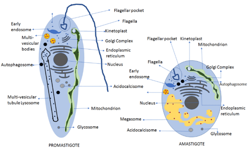
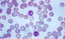
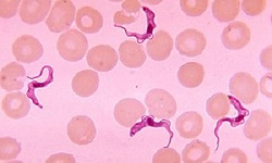
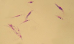
Figure: L. donovani morphology (Source: ResearchGate)
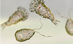
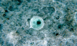
Morphology of Leishmania donovani
The protozoa parasites, Leishmania donovani, exist in two morphological forms- amastigote and promastigote form
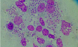
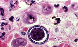
Amastigote form
amastigote form is found in humans and other mammal hosts
small, round to oval bodies, also known as Leishman Donovan (LD) bodies
each amastigote form measures 2-3μm in length
resides inside monocytes, endothelial cells, polymorphonuclear leucocytes of the host
the nucleus is large and lies at a right angle to the kinetoplast which is slender, rod-shaped
Leishmania donovani axoneme arises from the kinetoplast and extends to the margin of the amastigote
vacuole, which is not stained lies alongside the axoneme
stained well in Giemsa and Wright stain
in Giemsa-stained preparations, the nucleus, and kinetoplast, are stained red
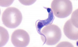
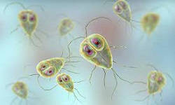
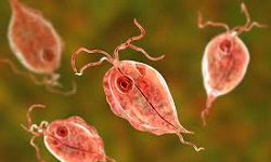
Promastigote
promastigote form occur naturally in the digestive tract of sand fly (vector)
mature promastigotes are long, slender, and spindle-shaped
measures 15 μm to 25 μm in length and 1,5 μm to 3.5 μm in breadth
a single nucleus is centrally located
kinetoplast located transversally near the anterior end
the presence of a single flagellum measuring 15μm -28 μm
Leishman stain stains the cytoplasm blue, the nucleus pink, and the kinetoplast bright red
Culture of Leishmania donovani
The culture of Leishmania donovani can be done in biphasic media, liquid media, or laboratory animals.
Biphasic media
consists of two parts of salt agar and one part defibrinated rabbit blood
Novy and McNeal (1904) and Nicolle (1908) (NNN) medium was the first biphasic medium for the culture of Leishmania donovani
Liquid media
includes Schneider’s, Grace’s, and Mituhasi-Maramorosh media- which are insect cell culture media
liquid media do not contain any blood
usually used for the preparation of a large volume of promastigotes
Schneider’s medium which contains 20% fetal calf serum (FCS) is more sensitive to culture than the NNN biphasic medium
Laboratory animals
Chinese hamsters and golden hamsters are routinely used in the culture and diagnosis of Leishmania donovani.