Loa loa - Culture, Life Cycle, Pathogenesis, Pathology, Host Immunity, Epidemiology, Reservoir, Source, Transmission
Culture of Loa loa
Loa loa has been cultured in laboratory animals by inoculation in Patas monkeys (Erythrocebus patas) and baboons (Papio anubis).
Life Cycle of Loa loa
The life cycle of Loa loa is completed in two hosts – the definitive host and the intermediate host.
Definitive host: Humans
Intermediate host: Female tabanid flies of genus Chrysops silica and Chrysops dimidiata
humans acquire Loa loa infection by the bite of female tabanid flies of genus Chrysops silica and Chrysops dimidiata during a blood meal
through the puncture wound on the skin, the mass number of infective larvae (L3) enter the subcutaneous tissue
inside the host tissue, Loa loa larvae molt, develop and sexually mature with a period of 6 to 12 months
as the adult worms migrate through the sub-conjunctival tissue and the subcutaneous tissue, they produce characteristic Calabar swellings
gravid females, which are viviparous, release microfilariae
these Loa loa microfilariae circulate in the peripheral blood during the day and move to the vascular parts of the lungs at the night
the microfilariae may also reside in the subcutaneous tissues of the host
when female tabanid flies of the genus Chrysops silica and Chrysops dimidiata take a blood meal, the Loa loa microfilariae are also picked up
inside the vector’s gut, the parasitic microfilariae lose their sheath and penetrate the stomach wall to reach the hemocoel and into the thoracic wing muscle
in the thoracic wing muscle, the filarial nematode parasite develops from a first-stage larva, second-stage larva, and finally into infective third-stage larvae (L3) in about 10 days
the infective larvae (L3) migrate to the proboscis (mouth parts) of the female tabanid flies
when the infected female tabanid flies take another blood meal, the Loa loa L3 larvae are transmitted to a new human host
the life cycle of Loa loa is then continued
Loa loa life-cycle (Source: CDC)
Pathogenesis, Pathology of Loa loa
Microfilariae of the filarial nematode parasite Loa loa are not pathogenic and do not produce any significant pathological lesions in the infected host.
Since adult Loa loa resides in the subcutaneous tissues, they are pathogenic. Calabar swelling, which is a typical feature of the infection, is caused by migrating adult worms in the subcutaneous tissue. It occurs due to an allergic response to the migrating adult parasites.
Host Immunity of Loa loa
Host Immunity against Loa loa infection includes hypersensitivity reaction to migrating adult worms.
Epidemiology of Loa loa
Epidemiological studies have shown that Loa loa infection is restricted to Africa and is endemic across Central Africa – prevalent in rain forests of both West Africa and Central Africa.
Reservoir, Source of Loa loa
The infected humans are the only source and reservoir of Loa loa infection. No animals are known to be the reservoir of the filarial parasite.
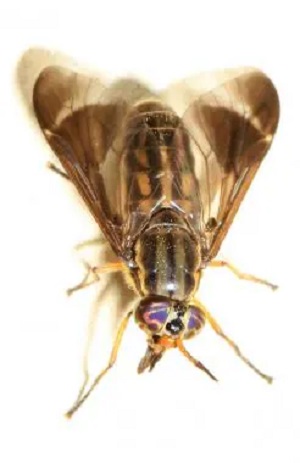
tabanid flies is a vector of Loa loa (Source: emedicine.medscape)
Transmission of Loa loa
Loa loa is transmitted man-to-man by the bite of female tabanid flies of the genus Chrysops silica and Chrysops dimidiata.