Loa loa - Introduction, Classification, History, Habitat, Morphology
Introduction to Loa loa
Loa loa is the etiological agent of the disease loiasis or Loa loa filariasis of sub conjunctiva of the eye and subcutaneous layer of the skin. The arthropod-borne filarial nematode, which is commonly referred to as eye worms, is commonly found in Africa. The roundworms cause transient swelling known as Calabar swellings.
Classification of Loa loa
The classification of Loa loa is as follows:
Kingdom: Animalia
Phylum: Nematoda
Class: Chromadorea
Order: Rhabditida
Family: Onchocercidae
Genus: Loa
Species: L. loa
History of Loa loa
Loa loa infection was first noted in the Caribbean in 1770 by Santo Domingo. Mongin, a French surgeon unsuccessfully tried to remove a worm passing across a woman's eye.
In 1778, surgeon François Guyot retrieved the parasite from a man’s eye, from a group of West African slaves on a French ship to America. Ophthalmologist Stephen McKenzie identified the Loa loa microfilariae while Calabar swellings were observed in 1895 by Scottish ophthalmologist Douglas Argyll-Robertson.
The cause-effect between Loa loa and Calabar swellings was linked in 1910 by Patrick Manson while the vector for the parasite was discovered to be Chrysops spp by British parasitologist Robert Thomson Leiper in 1912.
Habitat of Loa loa
Loa loa habitats the subcutaneous tissue of human hosts while unsheathed microfilariae are found in the blood.
Morphology of Loa loa
The body of Loa loa consists of the head, a body, and a blunt tail. The outer body contains a cuticle with 3 main layers which are made up of collagen. These later protect the parasite while they are inside the digestive system of their host.
Both adults and juveniles have similar morphological features and the only difference is size.
Significant morphological forms of Loa loa includes – adult worm, microfilaria, and the infective form (L3).
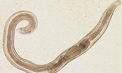
Adult worm
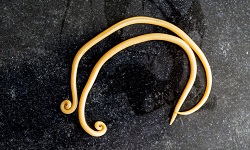
adult Loa loa worms are whitish, thun, thread-like
body surface covered with small knobs
anterior end tapers to a narrow head
resides in the subcutaneous tissue
travels under the host skin
life-span of 10 to 15 years
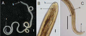
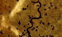
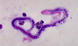
(A) Adult female Loa loa - head (h), tail (t); (B) lactophenol-cleared anterior extremity - (b) straight buccal canal, (o) muscular esophagus; (C) lactophenol- cleared posterior extremity -(a) anus, (u) posterior uterine loop of the posterior uterus (u) (Source: ResearchGate)
Male
male Loa loa measures 30 to 34mm in length and 0.35–0.42 mm in diameter
tail end has two spicules of unequal length
Female
female Loa loa measures 40–70 mm in length and 0.5 mm in diameter
vulva opens in the cervical region
deposits microfilariae under the host skin
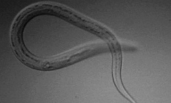
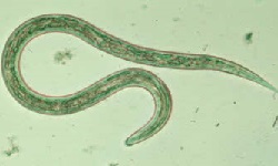
Microfilariae
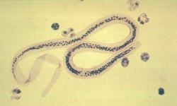
measures 250–300 μm in length, 6–8 μm in diameter
sheathed
sheath stains poorly or even remains unstained with Giemsa stain but stains well with iron-hematoxylin
contain body nuclei that extend to the tip of the tail
shows diurnal periodicity – appears in the peripheral blood during the day
in some cases, Loa loa microfilariae are found in the sputum, CSF, urine
can develop in the host’s blood within 5 to 6 months
survives upto 17 years
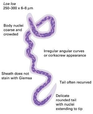
Loa loa microfilariae morphology (Source: Twitter)
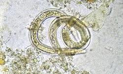
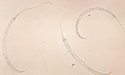
Infective form
The infective form of Loa loa is the stage-three larvae (L3). They develop in female tabanid flies (horseflies, deerflies) of the genus Chrysops silica and Chrysops dimidiata.
.jpg)