Paragonimus westermani - Introduction, Classification, History, Habitat, Morphology, Epidemiology, Reservoir, Transmission
Introduction to Paragonimus westermani
Paragonimus westermani is the etiological agent of pulmonary paragonimiasis or hemoptysis in humans. It is a lung fluke which is most common in eastern Asia and in South America.
Classification of Paragonimus westermani
Kingdom: Animalia
Phylum: Platyhelminthes
Class: Trematoda
Order: Plagiorchiida
Family: Paragonimidae
Genus: Paragonimus
Species: P. westermani
History of Paragonimus westermani
Coenraad Kerbert discovered Paragonimus westermani in 1878 in Bengal tigers kept in Amsterdam zoo. In 1879, the fluke was found during an autopsy of a Portuguese man while it was demonstrated by Manson in the sputum of a Chinese patient in 1880.
The life cycle of the parasite was eventually described by Yokogawa and Kobayashi between 1917 to 1921.
Habitat of Paragonimus westermani
In definitive hosts such as a human, adult Paragonimus westermani habitat in the parenchyma of the lung close to the bronchioles.
Morphology of Paragonimus westermani
The important morphological features of Paragonimus westermani include adult worms, eggs, and the infective form metacercaria.
Adult form
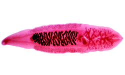
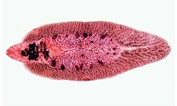
the adult form of Paragonimus westermani is thick and fleshy
egg-shaped fluke is reddish brown when freshly passed out
measures 7.5mm to 12mm in length, 4mm to 6mm in breadth, and 3.5 to 5mm in thickness
the anterior end is slightly broader than the posterior end
two muscular suckers – oral sucker and ventral sucker
* oral sucker is present in the anterior end
* ventral sucker is present on the ventral surface near the middle part of the body
the large excretory bladder which extends from the posterior extremity to the level of the pharynx
irregular deeply lobed testes are present side by side in the posterior end a characteristic feature of Paragonimus westermani
absence of cirrus sac
adult fluke has a lifespan of 6 to 7 years
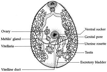
Image: Paragonimus westermani adult morphology (Source: Lecture Notes in Medical Technology)
Egg
Paragonimus westermani eggs are oval in shape, golden-brown in color
measures 48μm to 60μm by 80μm to 118μm
operculated eggs with clearly visible operculum at the large side and the opposite end without operculum are thick
unembryonated eggs are excreted in stool or sputum
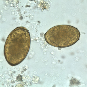
Image: Paragonimus westermani eggs (Source: CDC)
Infective form
The infective form of Paragonimus westermani is the metacercaria.
Metacercaria
infective to definitive host including man
encysted form is found in the flesh of crustaceans such as freshwater crayfish, crabs
Epidemiology of Paragonimus westermani
Epidemiology studies of Paragonimus westermani have confirmed infections in China, Japan, Korea, Taiwan, Laos, Thailand, Russian provinces, Peru, Ecuador, Philippines, India, etc.
Other species of Paragonimus have been reported in the Americas and Africa.
Reservoir, Source of Paragonimus westermani
The second intermediate host includes several genera of crab fish including Potamon, Sinopotamon, Parathelhusa and freshwater crayfish harboring metacercaria are the source of Paragonimus westermani infection.
The reservoir of Paragonimus westermani is the man, domestic animals, and wild carnivores. Body secretions such as sputum and stool containing Paragonimus westermani eggs contaminate water bodies such as lakes, pools, and rivers which infects the intermediate hosts freshwater snails belonging to the family Pleuroceridae, Thiaridae – Genera: Semisulcospira, Brotia while Semisulcospira libertina is the optimal intermediate host.
Transmission of Paragonimus westermani
Transmission of Paragonimus westermani occurs via the following routes:
consumption of raw, undercooked crab, and crayfish (also includes delicacy as soaked in wine or drinking raw crabs or crayfish)
less common – consumption of pork, wild pigs containing Paragonimus westermani larvae
via contaminated hands, utensils, and clothes used for staining