Plasmodium falciparum - Introduction, Classification, History, Habitat, Morphology, Culture
Introduction of Plasmodium falciparum
The genus Plasmodium causes the vector-borne disease malaria. These parasites show an alternation of generation accompanied by an alternation of hosts. In the human host, the asexual cycle (schizogony) takes place inside the erythrocytes while the sexual cycle (sporogony) takes place in the mosquito host.
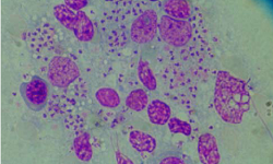
Typically, the infected erythrocytes produce pigments that are visualized by light microscopy.
Plasmodium vivax, P. ovale, and P. malariae belong to the subgenera Plasmodium while P. falciparum belongs to the subgenus Laverania.
Plasmodium falciparum causes falciparum malaria or malignant tertian and is the most virulent of Plasmodium species infecting humans. Almost all the serious forms of malaria, as well as the majority of malaria deaths, are caused by this parasite.
Classification of Plasmodium falciparum
Plasmodium falciparum can be classified by:
Kingdom: Chromista
Subkingdom: Harosa
Infrakingdom: Halvaria
Superphylum: Alveolata
Phylum: Apicomplexa
Class: Aconoidasida
Order: Haemospororida
Family: Plasmodiidae
Genus: Plasmodium
Species: P. falciparum
Habitat of Plasmodium falciparum
Plasmodium falciparum is found in various stages of malaria parasites in the human host. They habitats inside the parenchymal cells of the liver and the red blood cells.
Morphology of Plasmodium falciparum
The various morphological forms of Plasmodium falciparum depend on the host the parasite is residing in i.e. the diagnostic form or the infective form.
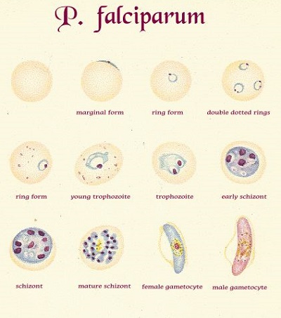
Fig: P. falciparum morphological forms (Source: Pinterest)
Diagnostic forms in humans
The diagnostic forms of Plasmodium falciparum found in the human host include
Early trophozoite (ring form)
Late trophozoite (trophozoite form)
Schizont
Gametocytes
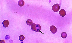
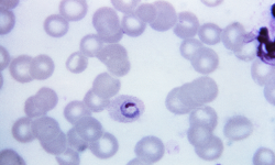
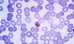
Early trophozoite (ring form)
sometimes, two early trophozoites can be found in a single infected RBC
are relatively large and rarely occur in peripheral blood
have a delicate blue-stained ring of cytoplasm with a red chromatin dot
in some cases, two red chromatin dots can be found separated or closed together
Late trophozoite (trophozoite form)
are amoeboid, vacuolated, uninucleated, and delicate
late trophozoite measures around 1.25μm to 1.5μm in size
in a stained preparation, a thin ring of blue cytoplasm and the darkish stained nucleus is present
the forms are compact in cases of heavy infections
yellow to black colored hemozoin, which are large masses of pigment, almost fill the RBC
hemozoin are insoluble polymers of haem which is an end product of ingested host hemoglobin
consists of abundant chromatin, dark pigment granules, and a vacuole which is characteristic
rarely seen in peripheral blood
Schizont
schizont are large, round, irregular, and asexual diving forms
occupy two third of infected RBCs, which have also been enlarged
hemozoin which almost fills the erythrocytes are present in one or two clumps
mature schizont contains around 10 to 36 merozoites in a grape-like cluster while the immature ones have only 2 to 4 merozoites
each merozoites measures 5μm to 10μm in length
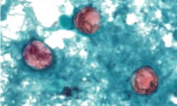
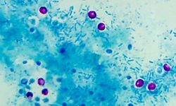
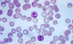
Gametocytes
Gametocytes are the sexual and erythrocytic stages of Plasmodium falciparum and are infective forms for mosquitos. They two types of gametocytes- macrogametocytes, and microgametocytes.
Macrogametocytes
Macrogametocytes measure 10μm in diameter
they are round or oval, compact, and filled with enlarged erythrocytes
smaller nucleus with a compact mass of chromatin
the fine granules are arranged in small masses and occur near the periphery of Plasmodium falciparum
the cytoplasm stains blue while chromatin pigments deep red and violet
Microgametocytes
microgametocytes are smaller than macrogametocytes
oval or round with a large nucleus
chromatin granules are arranged to form a spindle
cytoplasm, which is dark blue, contains dark, coarse hemozoin pigments distributed throughout the cytoplasm
do not occupy the entire host RBC
* Infected host erythrocytes
the infected host is not enlarged and contains several merozoites
‘applique’ and ‘accole’ forms are merozoites that lie along the red cell membrane
in late trophozoite forms, Maurer’s dots or clefts are common
*Infected host WBC
atypical mononuclear cells are observed
brown-black malaria pigments are found in monocytes and neutrophils
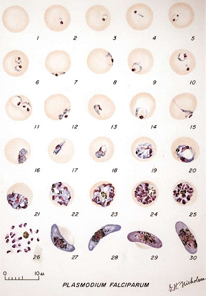
Figure: Developmental stage of P. falciparum - 1: Normal red cell; 2-6: Young trophozoites (ring stage parasites); 7-18: Trophozoites; 19-27: Schizonts; 28 and 29: Macrogametocytes (female); Microgametocyte (male) (Source: The Malarias Plasmodium falciparum - Welch 1898 )
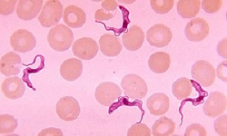
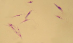
Infective form
The infective form of Plasmodium falciparum for humans is the sporozoites and is found in infected mosquitoes.
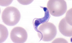
Sporozoites
sporozoites measures 10μm to 15μm
sickle-shaped with equally pointed ends and lack pigments
presence of a single nucleus
thick pellicle consists of a thin outer membrane, a two-layered membrane, and a layer of subpellicular microtubules
the posterior end has three polar rings and mitochondrion
organ of locomotion is the peripheral fibers
ookinete and oocyst are also found in the female Anopheline mosquito
found in the salivary gland of the mosquito
Culture of Plasmodium falciparum
The culture of Plasmodium falciparum can be done in-vitro or with laboratory animals.
In-vitro
The culture of Plasmodium falciparum is not done for routine diagnosis. It is rather used for
study the mechanism of invasion of erythrocytes by merozoites
biochemical and metabolic studies with each stage
source of antigen for serological as well as epidemiological studies
analysis of Plasmodium falciparum antigen
isolation of antigens for the development of vaccines
Plasmodium falciparum was cultured in-vitro for the first time in 1912 by Bass and Johns. Trager and Jensen cultivated and maintained Plasmodium falciparum in human RBC. This media has the following properties:
consists of RPMI 1940 medium
an overlay of medium with human serum
a thin layer of stationary human blood cells
maintained with 7% carbon dioxide in the atmosphere
maintained with 1% to 5% oxygen in the atmosphere
All the in-vitro methods to culture the parasite are modified versions of Trager and Jensen culture media.
Plasmodium falciparum is the only parasite widely cultivated in its asexual cycle.
Laboratory animals
The malaria parasite has been cultured in several species of laboratory animals such as primates such as monkeys.